Proanthocyanidins in seed coat’s tegmen and endospermic cap inhibit seed germination in the bioenergy plant Sapium sebiferum
- Published
- Accepted
- Subject Areas
- Agricultural Science, Plant Science
- Keywords
- Sapium sebiferum, seed dormancy, tegmen, endospermic cap, proanthocyanidins, ABA, GA
- Copyright
- © 2018 Shah et al.
- Licence
- This is an open access article distributed under the terms of the Creative Commons Attribution License, which permits unrestricted use, distribution, reproduction and adaptation in any medium and for any purpose provided that it is properly attributed. For attribution, the original author(s), title, publication source (PeerJ Preprints) and either DOI or URL of the article must be cited.
- Cite this article
- 2018. Proanthocyanidins in seed coat’s tegmen and endospermic cap inhibit seed germination in the bioenergy plant Sapium sebiferum. PeerJ Preprints 6:e3514v1 https://doi.org/10.7287/peerj.preprints.3514v1
Abstract
Sapium sebiferum, a highly ornamental and bioenergy plant, is propagated by seed. Its seed coat contains germination inhibitors and needs long time stratification for germination. In this experiment, we discovered that S. Sebiferum seed coat (especially tegmen) and endospermic cap contained high levels of proanthocyanidins (PAs). Seed coat and endospermic cap removal induced seed germination whereas exogenous application with seed coat extract (SCE) or PAs significantly inhibited this process, suggesting that PAs in the seed coat played a major role in regulating seed germination in S. sebiferum. We further investigated how seed coat extract affected the expression of the seed germination-related genes. The results showed that SCE treatment upregulated the transcription level of the dormancy-related gene, abscisic acid (ABA) biosynthesis and signalling genes and gibberellins (GA) suppressing genes. SCE decreased the transcript levels of ABA catabolic, GA biosynthesis, reactive oxygen species (ROS) and nitrates signalling genes. Exogenous application of nordihydroguaiaretic acid (NDGA), gibberellic acid (GA3), hydrogen peroxide (H2O2) and potassium nitrate (KNO3) recovered seed germination in SCE supplemented medium. In this experiment, we highlighted the role of PAs, and its interactions with the other germination regulators, in the regulation of seed dormancy in S. Sebiferum.
Author Comment
This is a submission to PeerJ for review.
Supplemental Information
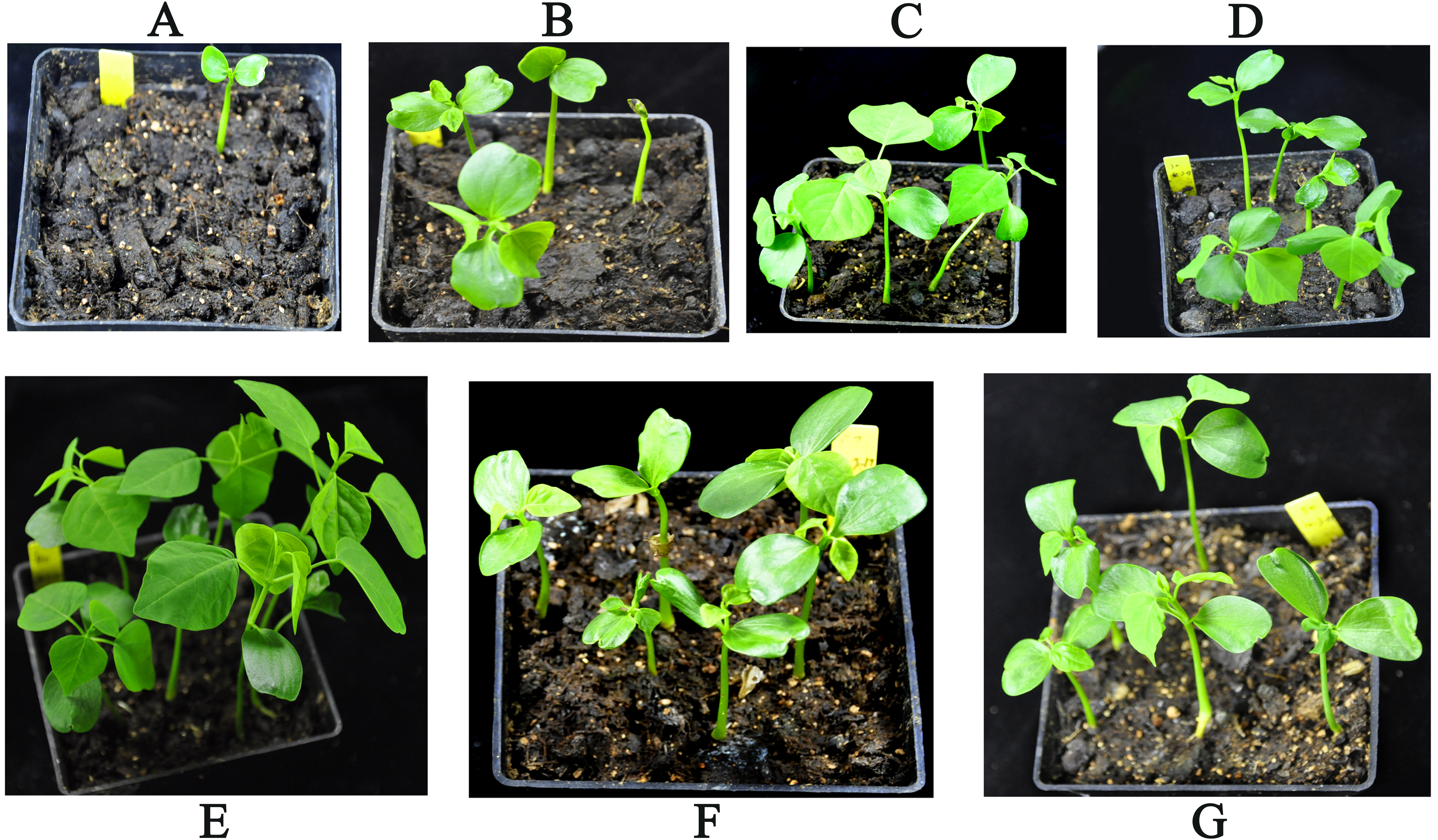
Impact of sulfuric acid stratification on seed germination of Sapium sebiferum
A, B, C, D, E, F and G are 0-(control), 10-, 20-, 30-, 40-, 50- and 60 minutes incubation in concentrated sulfuric acid respectively. Ten seeds of every treatment were sown in each 10×10 cm pot with 5 replicates. Photographs were taken after one month of seed sowing.
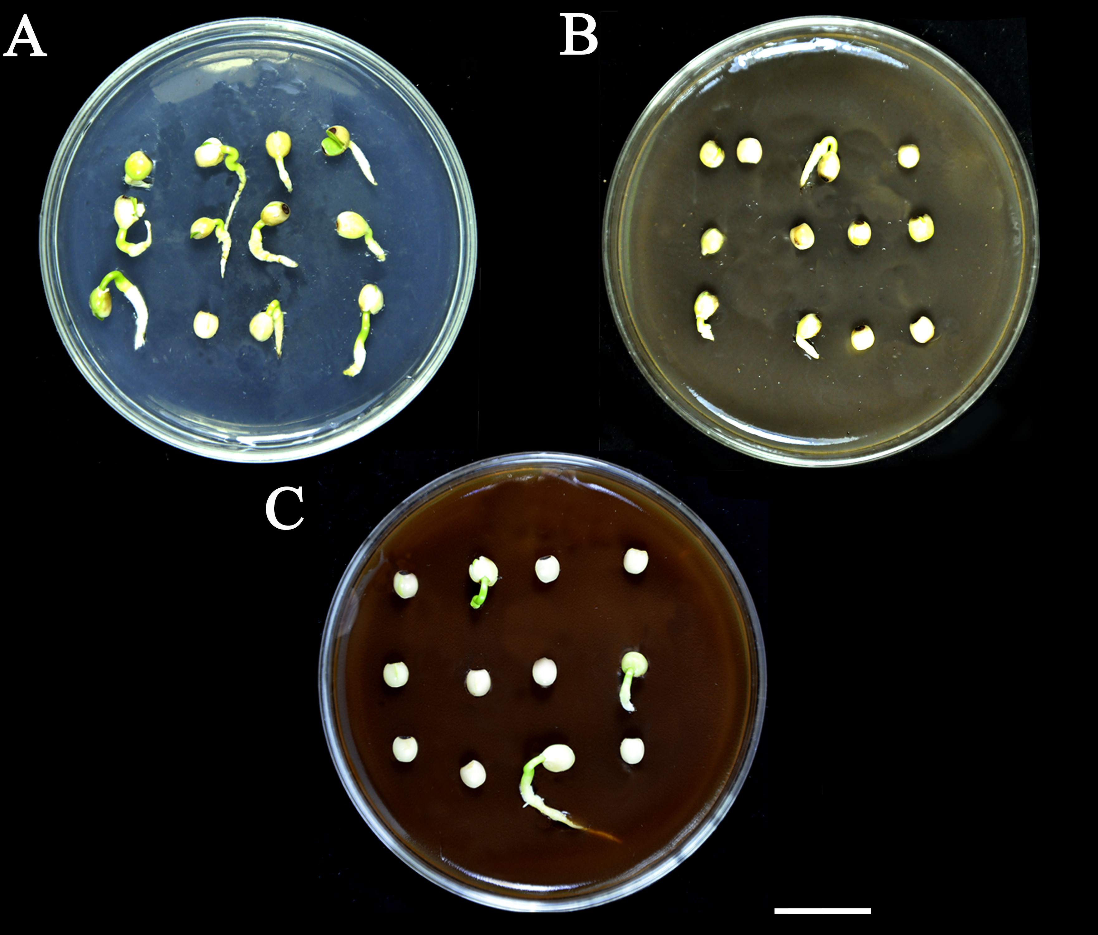
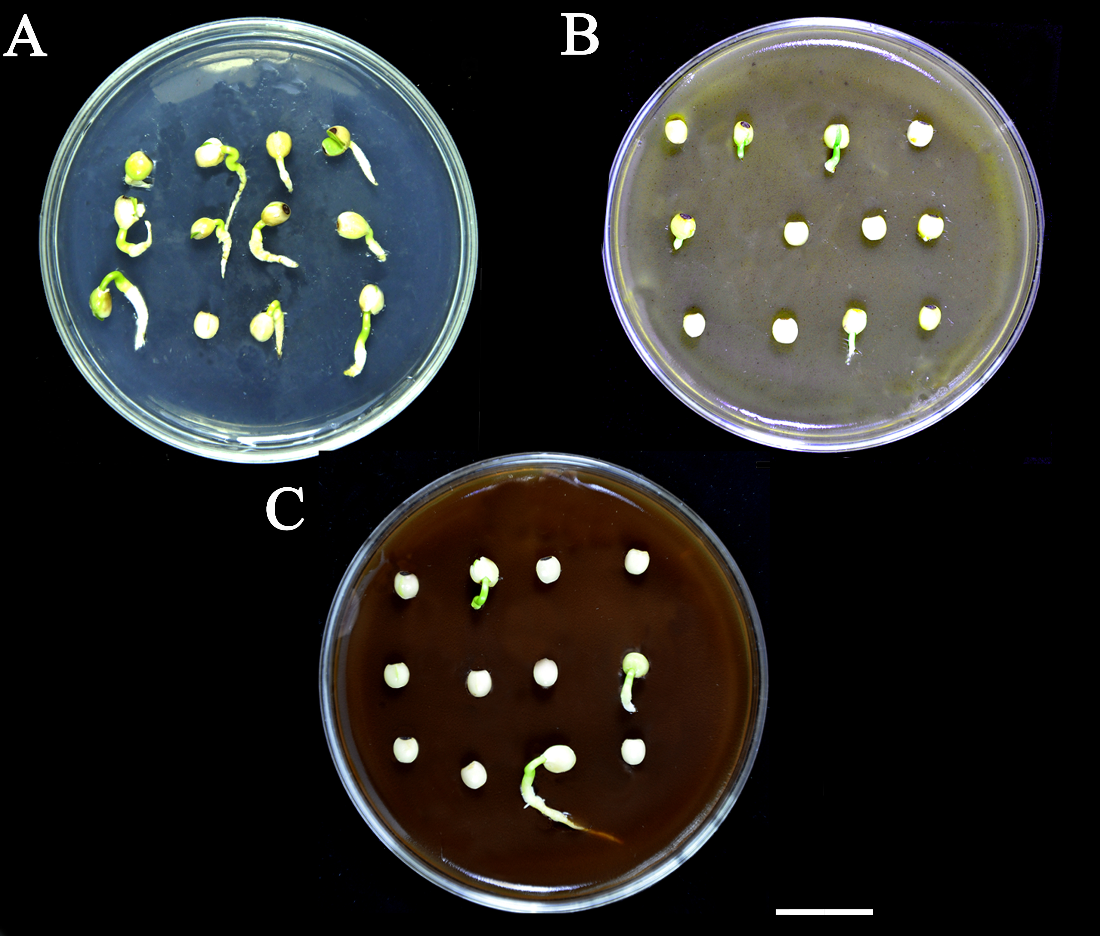
Effect of SCE and PAs on seed germination of S. Sebiferum
A, 0.5×MS (control). B, 0.3% SCE+0.5×MS. C, 0.1% PAs+0.5×MS. For each treatment, twelve seeds were sown in a 9 diameter cm Petri plate separately. All treatments were replicated 5 times. The Photographs were taken on the 7th day of imbibition. Bar = 2cm
Germination promoters recovered the seed germination sown in half MS supplemented with seed coat extract
A, control (sterile water). B, 50 μΜ GA3. C, 50 μΜ NDGA. D, 20 mM H2O2 and E, 0.4% KNO3 priming overnight at room temperature and the primed seed of all treatments were grown separately on 0.3% SCE+0.5×MS in 9 cm Petri plates (12 seeds per plate) for 7 days. These photographs were taken on the 7th day of imbibition. Bars = 1 cm >
water uptake percentage
Time, Scarification time. WUT, water uptake(%). Data was take 72 hours after imibibition
Effect of H2SO4 scarification time on seed germination
Treatment, H2SO4 scarification time. Germination, germination(%)
Root and shoot length of seedlings produced from H2SO4 scarified seed
Time, H2SO4 scarification time. Root, root length (cm). Shoot, shoot length (cm)
Effect of H2SO4 scarification time on PAs contents of seed coat
IT, H2SO4 scarification time. PAS, PAs contents (%) of seed coat after H2SO4 scarification
Datasheet of fig.5, 6 and 7
Data shows the delta C value of each gene. IDS, imbibition days
PAs contents in seed coat extract, testa and tegemen
PAs and SCE effect on seed germination
SCE and PAs were added in half MS. Data was taken after 7 days of imbibition
Data sheet of Figure 8 Impact of germination promoters
Germination, germination (%)