Modulatory interactions between the default mode network and task positive networks in resting-state
- Published
- Accepted
- Subject Areas
- Neuroscience
- Keywords
- dynamic connectivity, thalamus, salience network, basal ganglia, physiophysiological interaction, modulatory interaction
- Copyright
- © 2014 Di et al.
- Licence
- This is an open access article distributed under the terms of the Creative Commons Attribution License, which permits unrestricted use, distribution, and reproduction in any medium, provided the original author and source are credited.
- Cite this article
- 2014. Modulatory interactions between the default mode network and task positive networks in resting-state. PeerJ PrePrints 2:e124v2 https://doi.org/10.7287/peerj.preprints.124v2
Abstract
The two major brain networks, i.e. the default mode network (DMN) and the task positive network, typically reveal negative and variable connectivity in resting-state. In the present study, we examined whether the connectivity between the DMN and different components of the task positive network were modulated by other brain regions by using physiophysiological interaction (PPI) on resting-state functional magnetic resonance imaging data. Spatial independent component analysis was first conducted to identify components that represented networks of interest, including the anterior and posterior DMNs, salience, dorsal attention, left and right executive networks. PPI analysis was conducted between pairs of these networks to identify networks or regions that showed modulatory interactions with the two networks. Both network-wise and voxel-wise analyses revealed reciprocal positive modulatory interactions between the DMN, salience, and executive networks. Together with the anatomical properties of the salience network regions, the results suggest that the salience network may modulate the relationship between the DMN and executive networks. In addition, voxel-wise analysis demonstrated that the basal ganglia and thalamus positively interacted with the salience network and the dorsal attention network, and negatively interacted with the salience network and the DMN. The results demonstrated complex modulatory interactions among the DMNs and task positive networks in resting-state, and suggested that communications between these networks may be modulated by some critical brain structures such as the salience network, basal ganglia, and thalamus.
Author Comment
This manuscript was submitted for review with PeerJ.
Supplemental Information
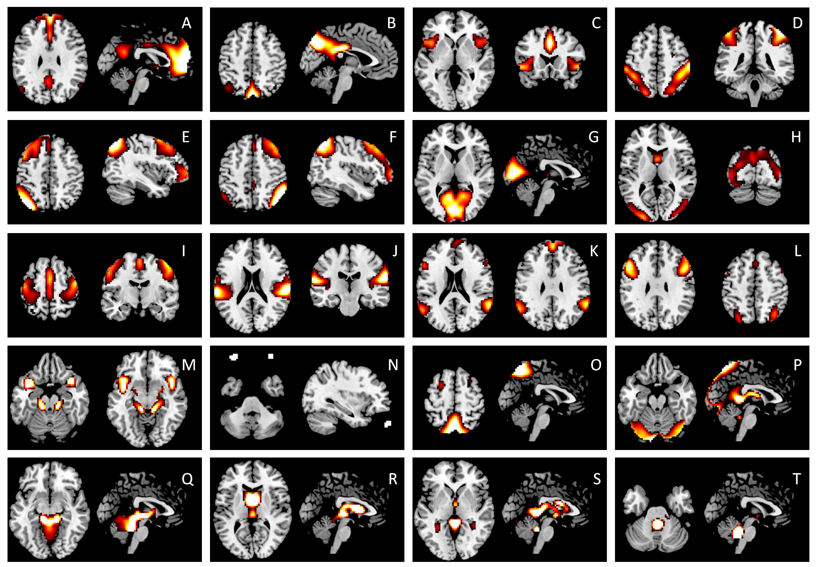
Full list of the 20 IC maps
The anterior DMN (A), posterior DMN (B), salience (C), dorsal attention (D), left executive (E), and right executive (F) components were used in the current PPI analysis. ICs G through J are categorized as unimodal networks, including the primary visual (G), higher visual (H), motor (I), and auditory (J) networks. ICs K and L are commonly identified components, however, their functions are not yet been clearly defined. We identified the remaining ICs (M through T) as noises, because their major clusters are located in either large blood vessels, ventricles, or eye balls. The IC maps were z transformed, and thresholded at z > 1.96.
Results of the network-wise PPI analysis
Numbers in each cell represent mean beta values and raw p values (in brackets) of corresponding cross subject one sample t-test. Bold font indicates statistically significant after Bonferroni multiple comparison correction of totally 60 correlations.
List of regions that showed significant PPI effects in voxel-wise analyses
Negative t values indicate negative PPI effects. Clusters were thresholded at p < 0.001, and cluster-level false discovery rate (FDR) correction was applied at p < 0.0033, which had taken into account of totally 15 voxel-wise analyses.