

Danielle performing immunohistochemistry in the lab. Photo credit: Ryan Thummel
Congratulations go to Danielle Quallich, winner of the PeerJ Award for Best Overall Presentation at the inaugural Ohio Zebrafish Undergraduate Research Conference. The conference was held at Ashland University on March 23, 2019. She presented her current research that adds to the growing body of work aimed at elucidating how adult zebrafish can regenerate their retinas following damage. This research has the long-term goal of generating new therapies and approaches to treat retinal pathology in human retinal degenerative diseases. The PeerJ Award, aimed at benefitting students and early career researchers, includes a free publication in PeerJ upon submission and acceptance through our normal peer review system. We are very pleased to recognize Danielle for her excellence in science and research.
![]()

Danielle receiving the PeerJ Award for Best Overall Presentation from Dr. Sarah Petersen, co-conference organizer along with Mason Posner. Photo credit: Mason Posner
-
Can you tell us a bit about yourself and your research interests?
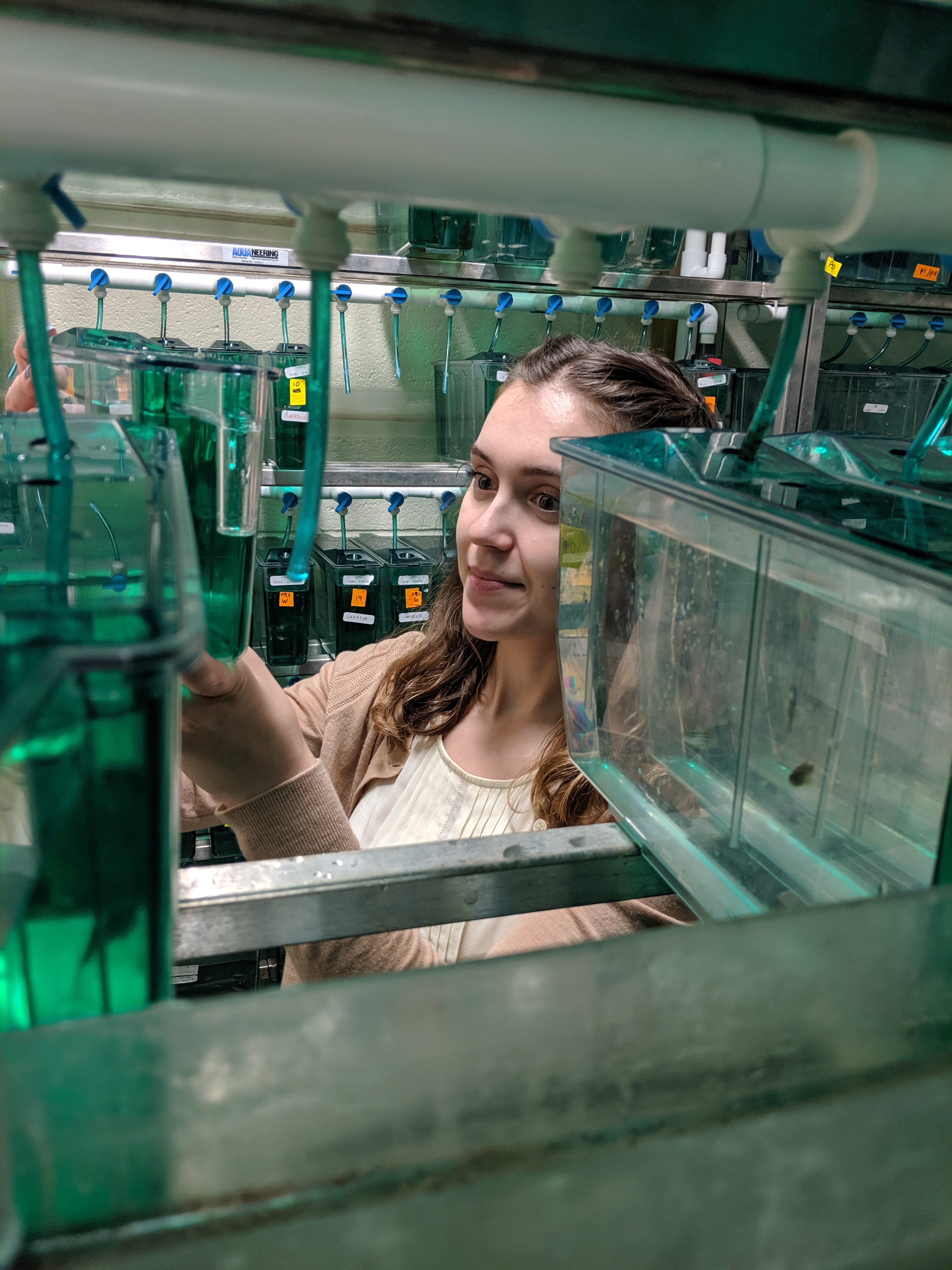
I attend Wayne State University in the Irvin D. Reid Honors College. My coursework has provided a strong background in multiple fields of science that aid my technical knowledge in a research environment. I’m an undergraduate research assistant in Dr. Ryan Thummel’s laboratory within the School of Medicine, Department of Ophthalmology, Visual and Anatomical Sciences. For the past two years, my work there has revolved around caring for the fish themselves and gaining experience with tissue harvesting, processing cryosectioning, RNA work, and immunohistochemistry staining. These methods have aided my experimental studies exploring a zebrafish model of retinal regeneration.

Feeding time for zebrafish. Photo credit: Ryan Thummel
I played a pivotal role in helping to develop a new chronic light-damage model to induce retinal neuron damage to observe the Müller glia proliferation response. Thus far, I have been responsible for performing the respective light treatments, the corresponding eye and retinal dissections and helping to compare and contrast the data from various time points. This chronic light-damage model was developed as a way to compare the stem cell response to that of a more acute, high-intensity light-damage model. It is imperative that these different light-damage techniques are compared so that the correlation between retinal damage and the stem cell response can be studied.
This area of research of is of specific interest to me as a chronic damage model will be more reflective of the slow loss of retinal neurons that occur in the majority of the retinal degenerative diseases in humans. Pursuing this research further would allow me to more actively study how specific cell layers in the eye and retina are affected by chronic and acute light-damage, leading to initiation of the stem cell proliferation response.
I have pursued my undergraduate studies while simultaneously serving the community of Detroit through volunteer opportunities. The more acquainted I become with diverse fields in the sciences, the more I have been drawn to the science of medicine. I have sought out community service projects that have directly complimented my course work, and environments that have exposed me to the fundamentals of patient care and family interaction. For example, volunteering at the Detroit Medical Center’s Children Hospital of Michigan has presented me with opportunities not only to analyze scientific and medical aspects of a situation, but that also require me to recall psychological and sociological themes in order to better engage with the children and their families.

Screening zebrafish embryos. Photo credit: Ryan Thummel
-
Can you briefly explain the research you presented at the Ohio Zebrafish Undergraduate Research Conference?
Many structural, functional and developmental similarities exist between zebrafish and human retinas, providing researchers with an excellent model to study human vision. Damage in mammals usually induces a scar-like response initiated by Müller glia, which aids in healing the affected area, but can prevent cells from returning to their original capabilities. In contrast, Müller glia in the zebrafish retina possess stem cell properties that allow them to respond differently to retinal damage. Specifically, zebrafish Müller glia proliferate to produce large numbers of retinal progenitor cells that ultimately replace lost retinal neurons and allow for full restoration to visual function. This remarkable regeneration event has been extensively studied as a reaction to various acute and severe damage events, such as exposure to high-intensity light. However, these damage paradigms fail to model most cases of retinal disease in humans, such as macular degeneration, which result in a slow loss of retinal neurons and vision. Therefore, the goal of this work was to develop a chronic low light-damage model in zebrafish in order to compare the stem cell response of resident Müller glial cells to that of an established, more acute, high-intensity light-damage model.
The effects of a low-intensity light treatment were mainly unknown prior to this experiment which is why the same timepoints post the initiation of the low-light treatment were used for each staining procedure and primary antibody immunolabeling. This was done to study the effects of the chronic model in a more comprehensive way. Post the initiation of a high-intensity light paradigm, data indicates that apoptosis, an initiation of a cascade of responses to being specified cell death without initiating an inflammatory response, was commonly seen when acute damage has occurred to the zebrafish retina. This study investigated the possibility of apoptosis in response to the low-intensity light with TUNEL analysis; a special technique that allows for the observation of cells undergoing apoptosis. The data collected in this study did not present with evidence of apoptosis, however, damage was seen in the low-light treated zebrafish retinas as evidenced by the fact that rod photoreceptor cells were present in fewer and fewer quantities throughout the duration of the experiment. Zpr-3 immunolabeling of rod photoreceptors indicated that a low-intensity light treatment facilitates slow degeneration of rod outer segments in the absence of apoptotic cell death.
Danielle observing zebrafish in the fish room. Photo credit: Ryan Thummel
Previous literature has concluded that the Müller glia residing within the zebrafish retina are the major source of proliferating progenitor cells following acute retinal damage. In contrast, data collected from the present study show no evidence of Müller glia proliferation (Thummel et. al, 2008; Thomas & Thummel, 2013). Müller glia projections, immunolabeled with glutamine synthetase, were neither hypertrophied nor did the Müller glia produce large quantities of proliferating progenitors typically seen with a Müller glia regeneration response. The lack of a Müller glia proliferation response contrasted the rod precursor proliferation seen as a response to the low-intensity light treatment. PCNA was used to immunolabel cells that were reentering the cell cycle; as the low-light treatment progressed, rod precursor cell proliferation occurred in the outer nuclear layer. As rod precursor cells are influential to the growth of rod photoreceptors and help mediate retinal cell regeneration in response to damage, these data indicated that the damage induced by the low-intensity light treatment was not enough to evoke the Müller glia proliferative regeneration response, but, sufficient damage was done to induce rod precursor cells to reenter the cell cycle.
This study showed that a low-intensity light paradigm evoked retinal degeneration was occurring in the absence of apoptosis and the central nervous system seemed to respond via its macrophages. The primary antibody 4C4 tags microglial cells, macrophages of the central nervous system, that engulf large debris to package and dispose it via its lysosomes. Microglia reside within the inner and outer plexiform layers and as the time of the treatment progressed, more and more microglia presented in an attempt to clear away the debris of degrading cells. This indicated that, while no apoptosis was occurring, damage was still induced by the low-light treatment to the point where the central nervous system of zebrafish require its microglia to clear cellular debris from the rod out segments and the outer nuclear layer. Overall, this research adds to the growing body of work aimed at elucidating how adult zebrafish can regenerate their retinas following damage and has the long-term goal of generating new therapies and approaches to treat retinal pathology in human retinal degenerative diseases.
-
What are your next steps? How will you continue to build on this research?
To add to the data of this low-intensity light treatment, further antibody related immunohistochemistry procedures could be done on zebrafish tissues. For example, zebrafish also contain blue and ultraviolet photoreceptors and antibodies exist to immunolabel such cells. Further studies could investigate the fate of these types of cone photoreceptors within the zebrafish retina in response to a chronic light treatment. Long term goals would be the exploration of which genes are activated in the acute model and to compare those genes to the genes activated in the chronic model. The Müller glia proliferation response leads to stem cell regeneration in zebrafish and a scar tissue response in humans; an experiment could be designed to study the genes that are activated in the zebrafish in response to damage that ultimately lead to Müller glia stem cell mechanism. These genes and gene pathways could be studied in comparison to the genes that are expressed in humans and to possibly discover if a similar gene in humans exists.
View PeerJ research in developmental biology, molecular biology, neuroscience, and ophthalmology. Read up on additional zebrafish research from Dr. Thummel’s lab: Characterization of retinal regeneration in adult zebrafish following multiple rounds of phototoxic lesion and also from Mason Posner et al.: The zebrafish as a model system for analyzing mammalian and native α-crystallin promoter function.
About: PeerJ is an Open Access publisher of seven peer-reviewed journals and a preprint server. PeerJ’s mission is to help the world efficiently publish its knowledge. All works published by PeerJ are Open Access and published using a Creative Commons license (CC-BY 4.0).
PeerJ – the Journal of Life and Environmental Sciences is the peer-reviewed journal for Biology, Medicine and Environmental Sciences. PeerJ also publishes PeerJ Computer Science, and five newly launched PeerJ Chemistry journals.
By teaming up with a number of conferences to offer these awards, we are making it as easy as possible for organizers to reward excellence in science, support students and early career researchers, and signal to the wider research community that open science is better science. Learn more here and get in touch if you are a conference organizer and are looking to offer a ‘Best Contribution’ award for open science – [email protected]