A novel animal model for neuroinflammation and white matter degeneration
- Published
- Accepted
- Subject Areas
- Neuroscience, Neurology, Pathology
- Keywords
- shRNA, Drd1, microglial activation, innate immunity, neuroinflammation
- Copyright
- © 2017 Ji et al.
- Licence
- This is an open access article distributed under the terms of the Creative Commons Attribution License, which permits unrestricted use, distribution, reproduction and adaptation in any medium and for any purpose provided that it is properly attributed. For attribution, the original author(s), title, publication source (PeerJ Preprints) and either DOI or URL of the article must be cited.
- Cite this article
- 2017. A novel animal model for neuroinflammation and white matter degeneration. PeerJ Preprints 5:e3117v1 https://doi.org/10.7287/peerj.preprints.3117v1
Abstract
Small interference RNA has been widely used to suppress gene expression. Three different short-hairpin RNAs (shRNAs) against dopamine D1 receptor (Drd1), driven by mouse U6 promoter in self-complementary AAV8 vector (scAAV8), were used to silence mouse striatal Drd1 expression. Transduction of mouse striatum with all 3 scAAV8-D1shRNA virus, but not the control scAAV8 virus, causes extensive neuroinflammation, demyelination, and axon degeneration. RNA interference is known to be coupled to the innate immune system as a host cell defense against virus infection. Activation of the innate immune system may play a causal role in the development of neuroinflammation and white matter degeneration, providing a novel animal model for multiple sclerosis (MS) and other neuroinflammatory diseases.
Author Comment
This is a submission to PeerJ for review.
Supplemental Information
Design of D1shRNAs and suppression of Drd1 protein in mouse striatum
(A) Three different shRNAs were designed to target different sites of mouse Drd1 gene. Immunohistochemical staining of Drd1 was performed using rabbit anti-Drd1 antibody in mouse striatum transduced with either scAAV8-hrGFP (B) or scAAV8-D1shRNA1 (C) virus. The virus was injected into the left striatum only. Reduced Drd1 protein was observed in the left striatum injected with scAAV8-D1shRNA1 virus, but not with scAAV8-hrGFP virus. High magnification of Drd1 reduction was shown in the left striatum transduced with scAAV8-D1sdhRNA1 virus (D) in comparison with Drd1 expression in the right control striatum (E). Scale bar: 50 µm
Mild neuroinflammation in striatum transduced with scAAV8-D1shRNA2 virus
(A) Immunohistochemical staining of Iba-1 in mice injected with scAAV8-D1shRNA2 virus. Microglial cells were activated to surrounding blood capillaries in the left striatum transduced with the scAAV8-D1shRNA2 virus. (B) and (C) are the higher magnifications of mild neuroinflammation around the blood capillaries. Scale bar: 50 µm.
Immunohistochemical staining of MBP
There is no difference in MBP staining in the left corpus callosum transduced with scAAV8-hrGFP virus (A) in comparison with the control right corpus callosum without virus injection (B). Scale bar: 100 µm. MBP was reduced (black arrow) on the left corpus callosum (C) of mice injected with scAAV8-D1shRNA3 in comparison with the normal right corpus callosum (D). Scale bar: 200 µm. Under a higher magnification, MBP staining was also decreased in striatal white matter tracts that were swollen and blebbed (black arrowheads) in the left striatum transduced with scAAV8-D1shRNA3 (E) in comparison with normal white matter tracts in the control right striatum (F). Scale bar: 60 µm.
Absence of neuroinflammation and white matter degeneration in the left striatum transduced with scAAV8-hrGFP virus
Two consecutive paraffin sections from mice injected with scAAV8-hrGFP virus were immunostained 7 weeks post-injection with either anti-Iba-1 (A) or anti-NF-L (B) antibody. There was no activation of microglial cells in the left striatum transduced with scAAV8-hrGFP virus in comparison with the uninjected control right striatum. There was no reduction of NF-L staining in the corpus callosum on the left side (C) transduced with the virus in comparison with the control right side without virus infection (D). Scale bar: 150 µm.
White matter degeneration and neurofilament reduction in striatum transduced with scAAV8-D1shRNA3 virus
Two consecutive brain paraffin sections from mice injected with scAAV8-D1shRNA3 virus were immunostained with either anti-Iba-1 (A) or anti-NF-L (B) antibody. Extensive microglial activation as shown by anti-Iba-1 immunostaining was observed in the left striatum transduced with the scAAV8-D1shRNA3 virus in comparison with the control right striatum. Decreased NF-L staining was observed in the left corpus callosum (C) compared with the control right corpus callosum (D). Scale bar: 200 µm. Neurofilament staining was reduced in blebbed striatal white matter tracts (black arrow) in the left striatum transduced with the virus (E) in contrast to the control right striatum (F). Scale bar: 60 µm.
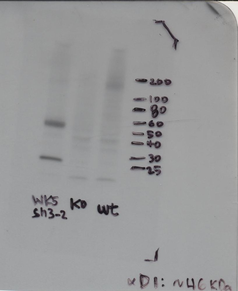
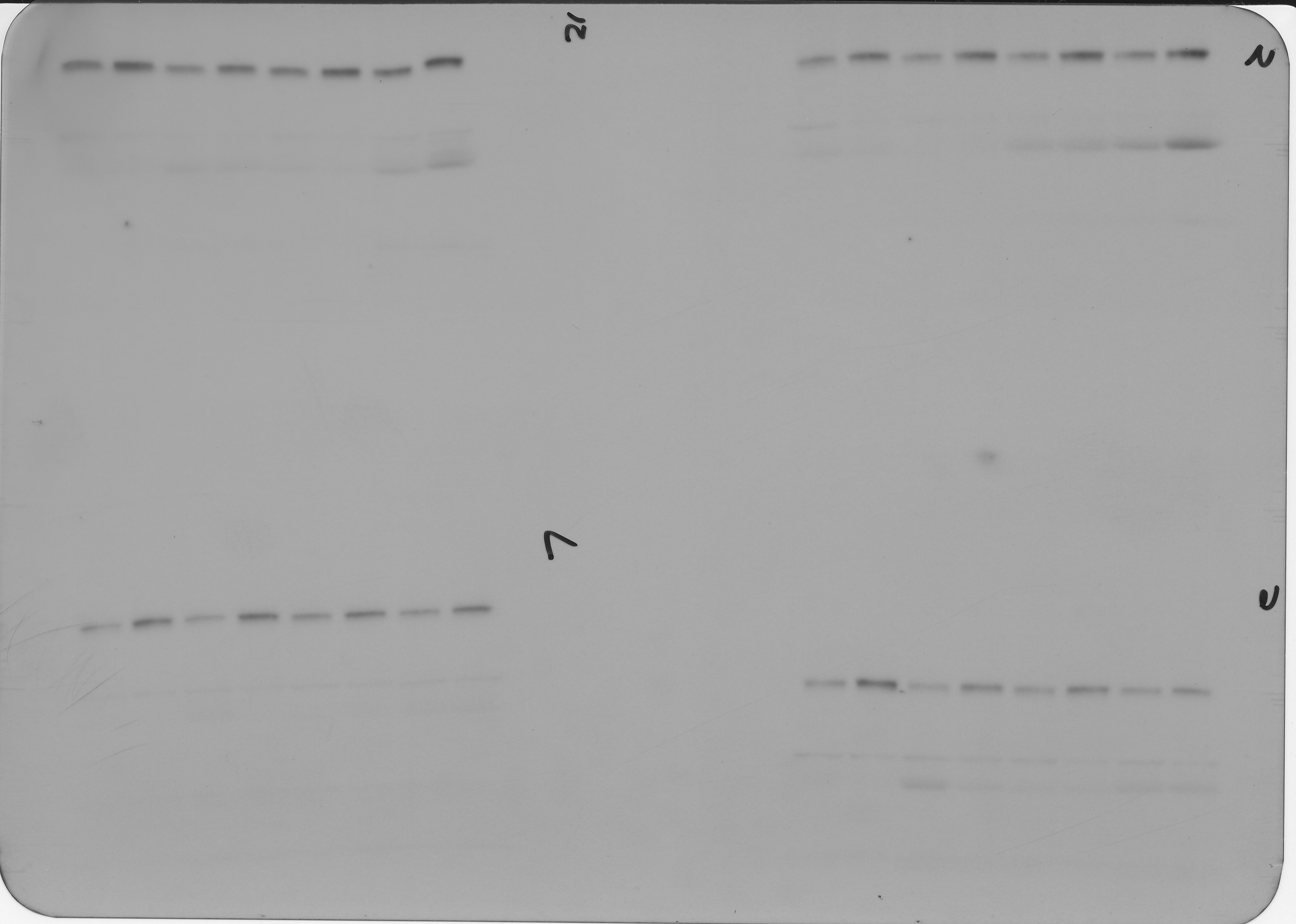
Western blot raw data mouse IgG week 7 to 15
week 7, 8, 9, 15
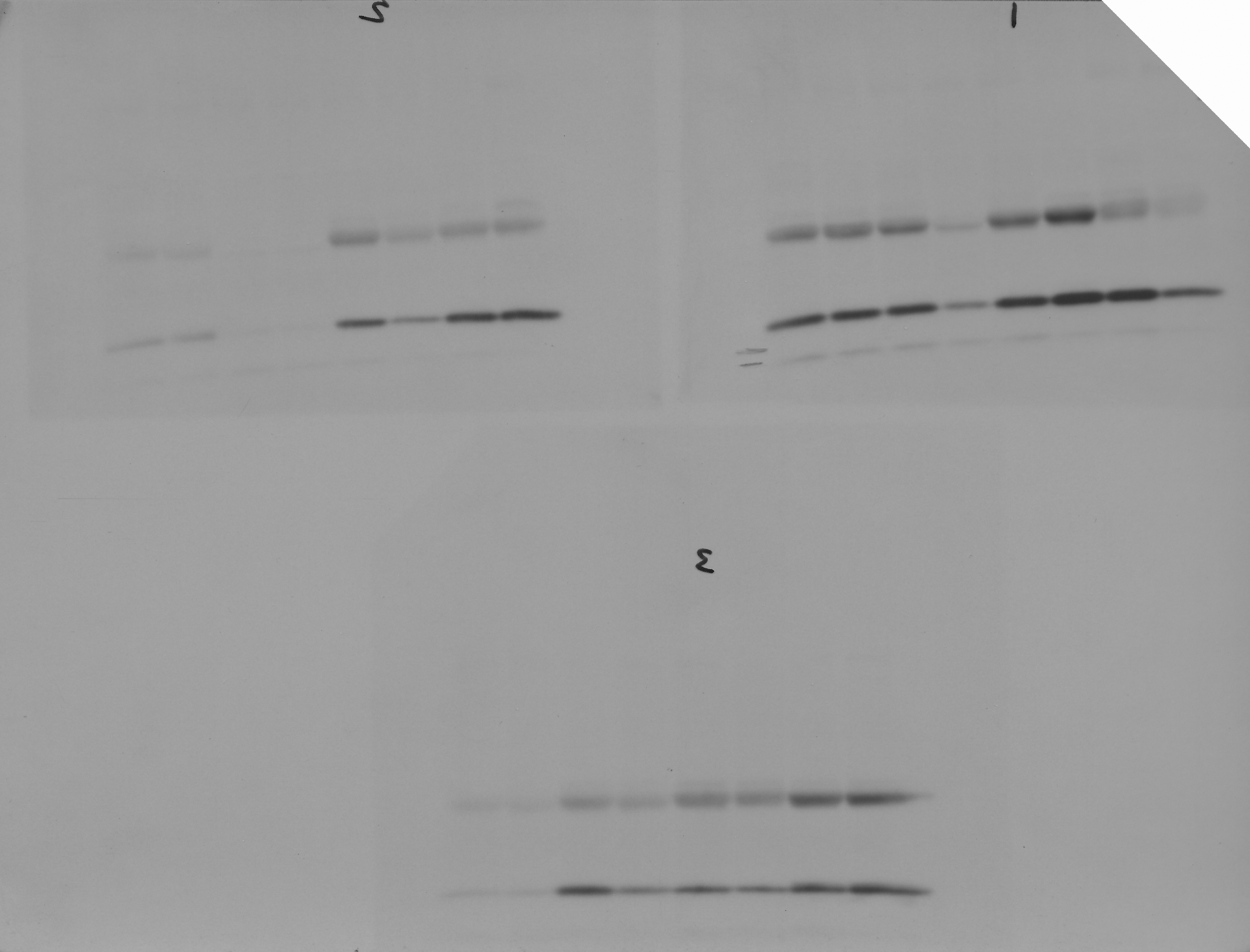
Western blot raw data NR1 week 4 to 6
normalization controls
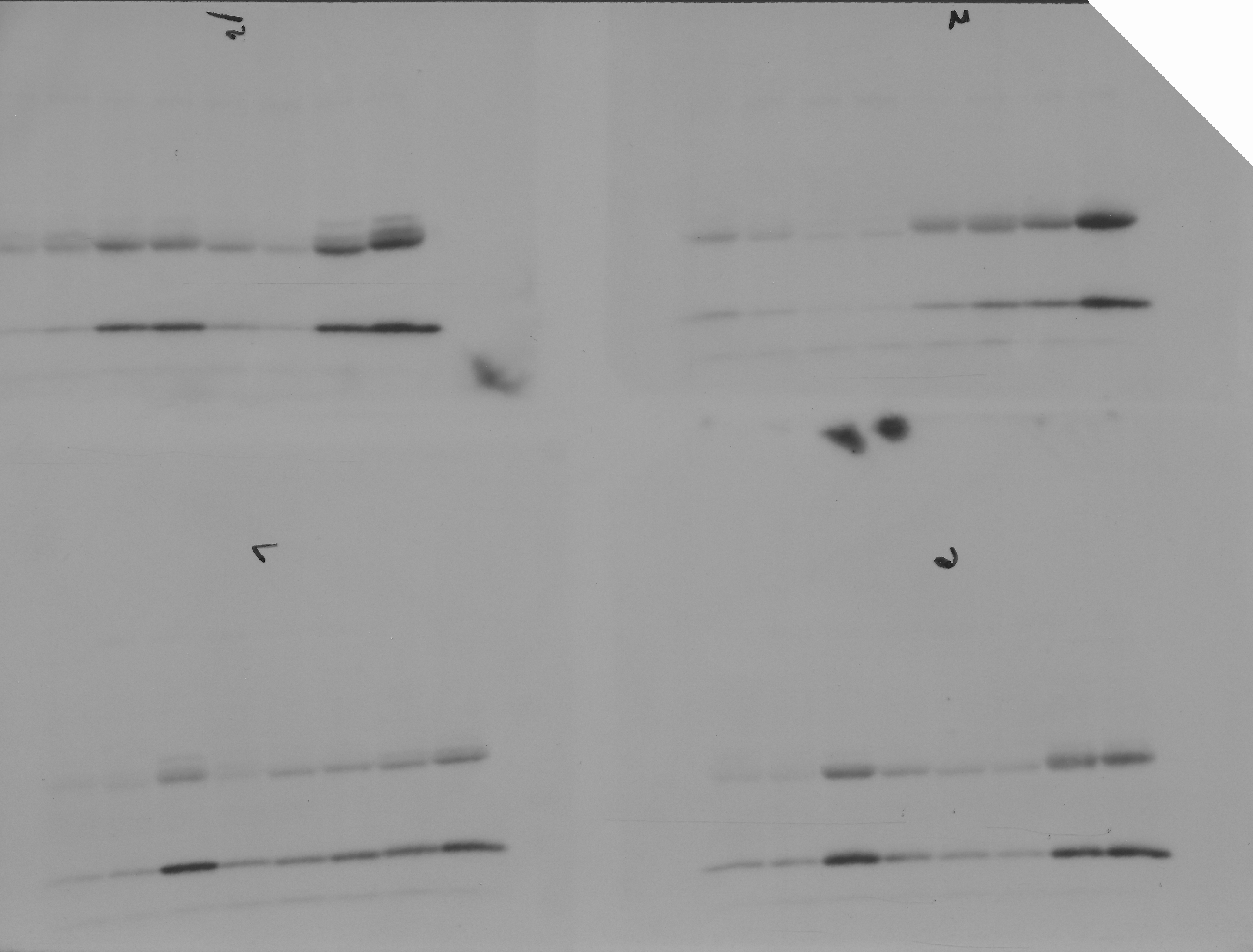
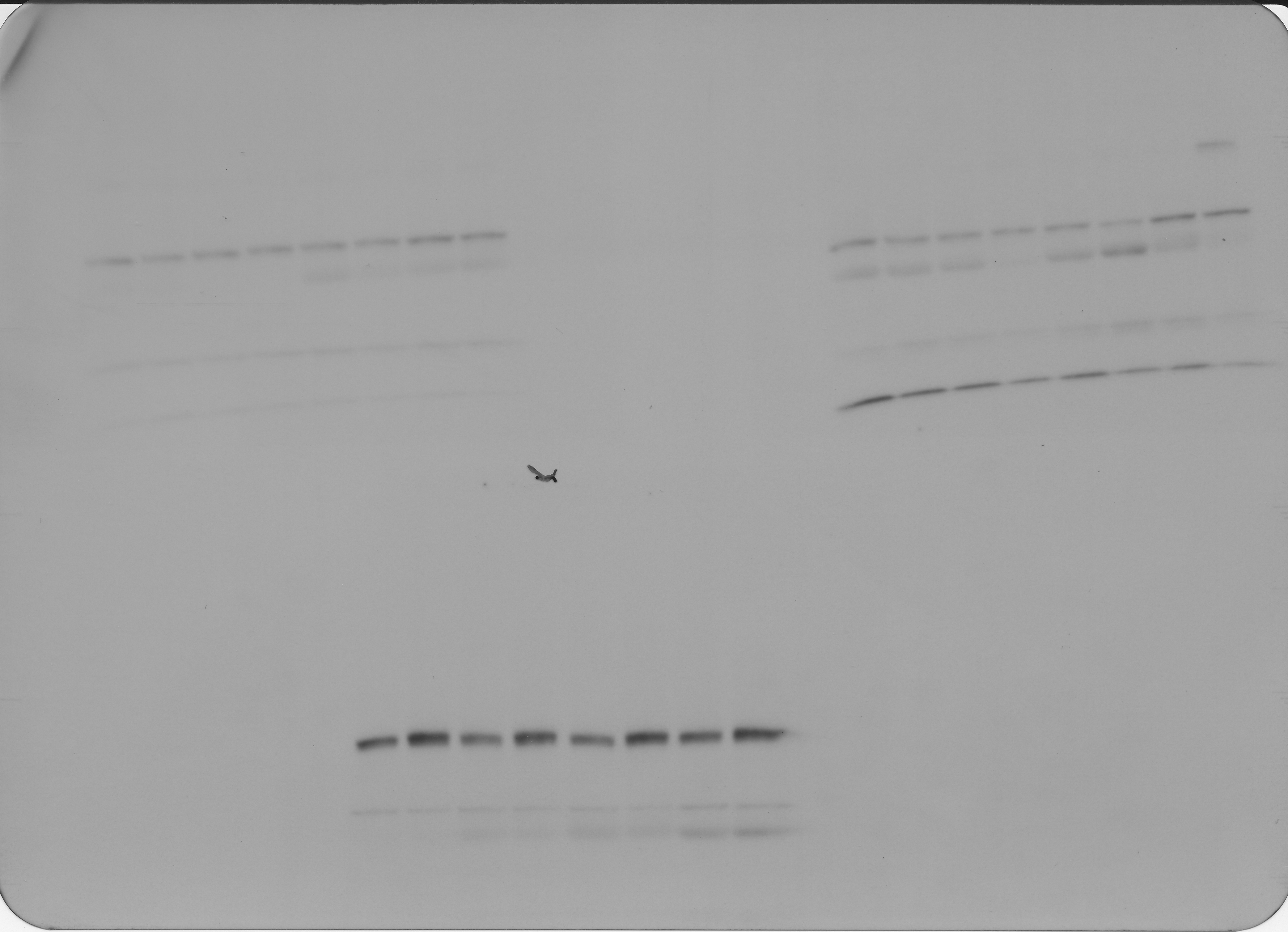
Western blot raw data NR1 week 7 to 15
Normalization controls for week 7,8,9, and 15.