Arabidopsis PEN2, a promising gene in upraising penetration resistance against rice necrotrophic fungus Rhizoctonia solani
- Published
- Accepted
- Subject Areas
- Agricultural Science, Cell Biology, Genetics, Plant Science
- Keywords
- necrotroph, Rhizoctonia solani, Nonhost resistance, infection cushion, penetration
- Copyright
- © 2019 Parween et al.
- Licence
- This is an open access article distributed under the terms of the Creative Commons Attribution License, which permits unrestricted use, distribution, reproduction and adaptation in any medium and for any purpose provided that it is properly attributed. For attribution, the original author(s), title, publication source (PeerJ Preprints) and either DOI or URL of the article must be cited.
- Cite this article
- 2019. Arabidopsis PEN2, a promising gene in upraising penetration resistance against rice necrotrophic fungus Rhizoctonia solani. PeerJ Preprints 7:e27611v1 https://doi.org/10.7287/peerj.preprints.27611v1
Abstract
Rhizoctonia solani, a soilborne necrotroph, causes sheath blight in rice which poses a major threat to global rice production. Besides rice sheath blight, it has a wide host range of other economically important crops like soybean, sugarcane, maize etc. Despite being the most hostile fungus, the mechanism involved in the R. solani pathobiology is poorly understood. Non-host resistance (NHR) is an emerging concept that allows breeders to transfer traits to food crops that would impart a broad-spectrum disease resistance. Several NHR genes are known to function against different pathogens of which Arabidopsis PEN1, PEN2 and PEN3 have been reported to limit the entry of non-adapted powdery mildews and provide cell wall based defenses against different fungi. Till now, there has been no study regarding the involvement of these PEN genes against R. solani. In this study, we have screened pen1, pen2-3 and pen3-1 against R. solani to explore their contribution in penetration resistance. Among the three pen mutants studied, pen2-3 allowed maximum penetration during the early hours of infection. R. solani colonization was also observed in pen1 and pen3-1 but the effect was less drastic than pen2-3, suggesting the involvement of PEN2 in pre-invasive defense. To validate our hypothesis, we screened a complemented pen2 accession, PEN2-GFP, which showed restored penetration resistance comparable to Col-0. Altogether, our results demonstrate that PEN2 is involved in pre-penetration resistance, and contributes to NHR by enhanced disease resistance to R. solani.
Author Comment
This is a submission to PeerJ for review.
Supplemental Information
PCR based confirmation of homozygous PENETRATION mutants used in the study
(a) the T-DNA insertion mutant of pen1 (SALK_004484C) was confirmed using a T-DNA left border primer indicating the presence of insert in the mutant. (b) pen2-3 was confirmed using a CAPS marker indicating the presence of an additional restriction site for BsmAI restriction enzyme. (c) pen3-1 was also confirmed using a CAPS marker showing deletion of HphI restriction site in the mutant.
Infection phenotypes of Arabidopsis wild type and pen mutants upon infection with R. solani
Leaves of Col-0 and pen mutants were stained with trypan blue after 6, 12, 24, 30, 36 and 48hpi to observe the extent of disease progression. Stained leaves were observed under bright field microscope. Leaves inoculated with water were used as control. Scale bar = 100µm
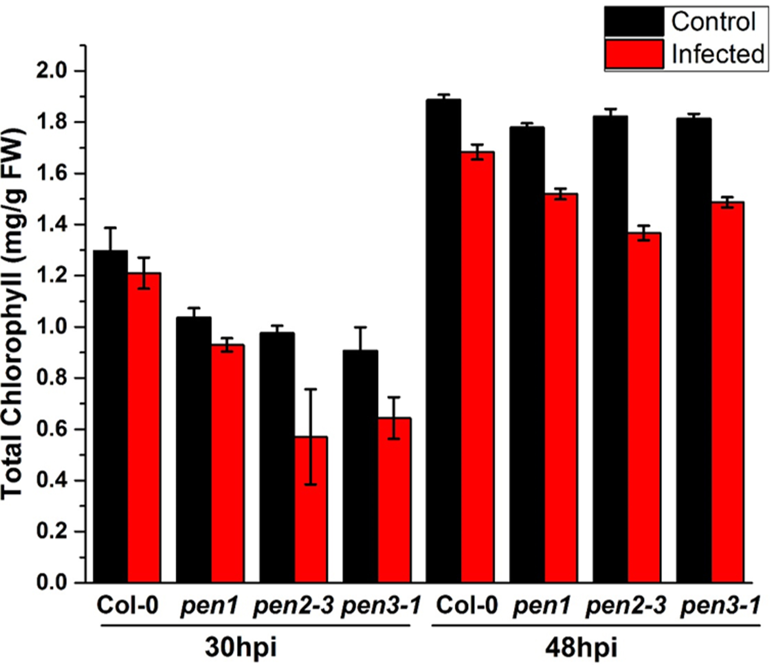
Total chlorophyll estimation upon infection with R. solani on Arabidopsis wild type (Col-0) and pen mutants
Total chlorophyll content measured in leaves of wild type and pen mutants of A. thaliana after 30 and 48hpi with R. solani sclerotia. Leaves treated with water served as control. Data represent the mean ± SEM of three biological replicates (n = 3). The experiment was carried out three times with similar results
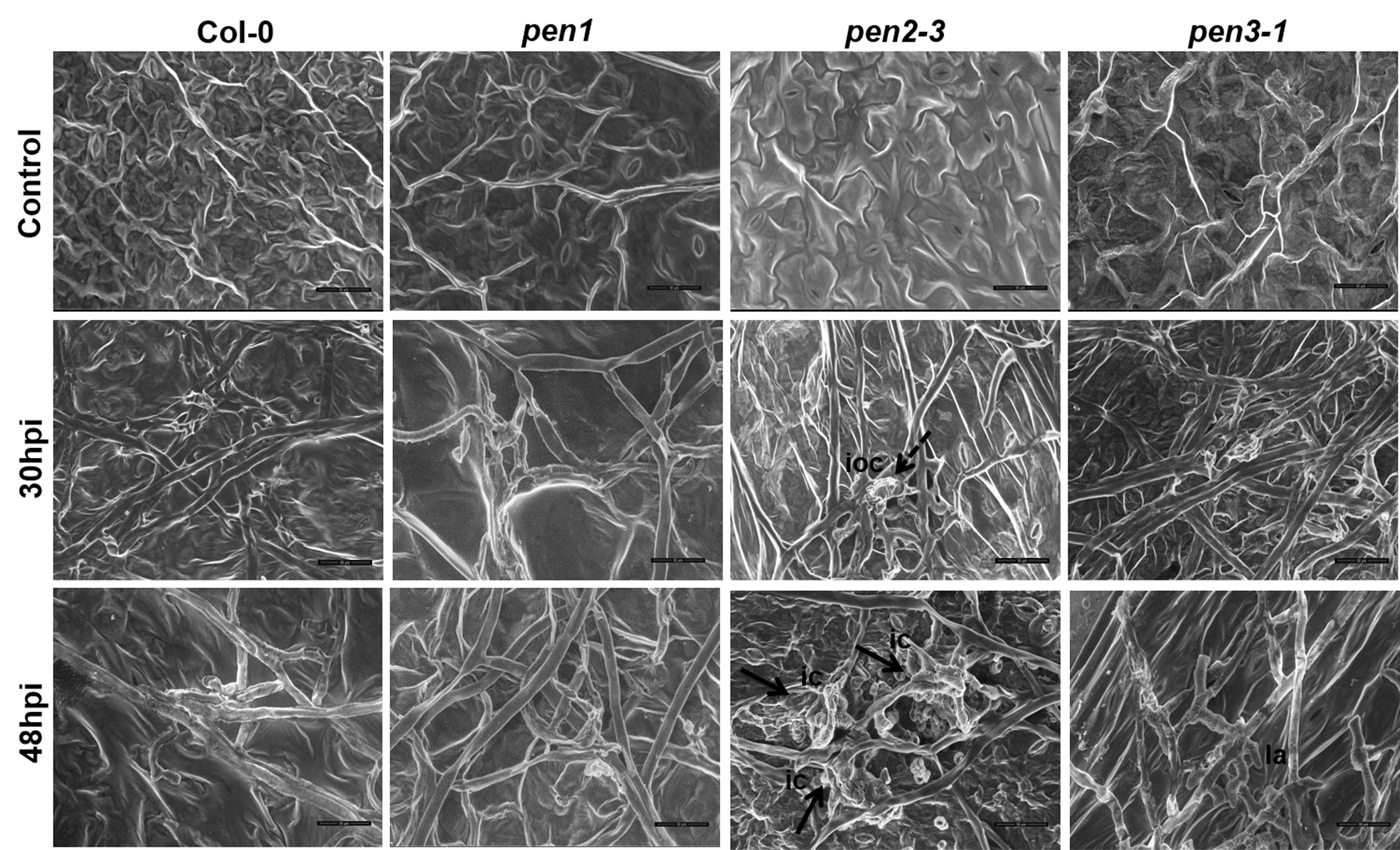
SEM micrographs of Arabidopsis pen mutants showing post infection hyphal colonization by R. solani at 30hpi and 48hpi
Leaves of Arabidopsis wild type (Col-0) and pen mutants inoculated with water as control. Col-0 and pen mutants inoculated with R. solani sclerotia showed disease progression with profuse hyphal branching after 30hpi. pen2-3 showed sporadic onset of infection cushion formation (ioc; dashed arrow) after 30hpi. Col-0 and pen mutants inoculated with R. solani sclerotia observed after 48hpi. Dense infection cushions (ic; solid arrows) at 48hpi in pen2-3. Swollen hyphal tip formed lobate apperesoria (la) at 48hpi in pen3-1. Scale bar ~ 50µm
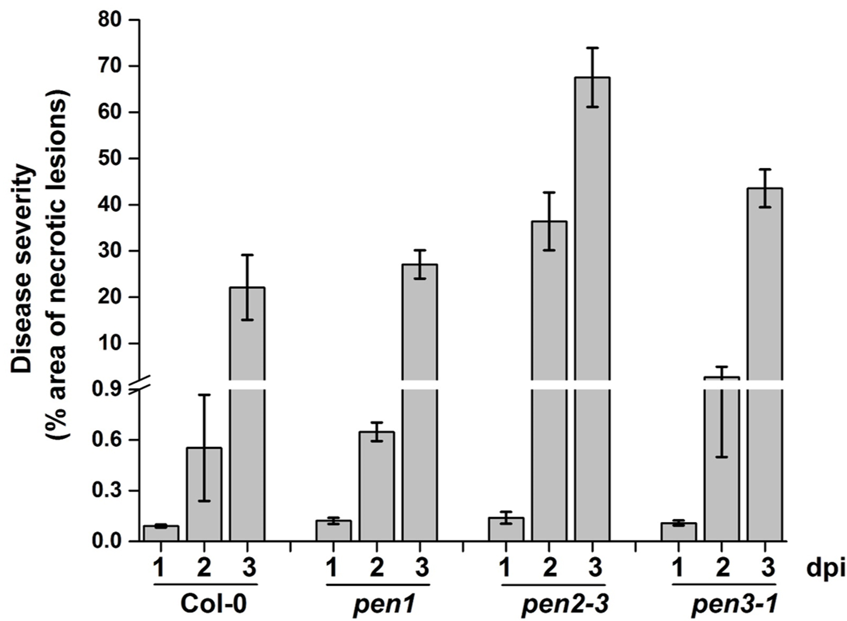
Macroscopic quantification of disease severity of R. solani infected Arabidopsis pen mutants
Macroscopic necrotic lesions were quantified in terms of area using ImageJ software after 1, 2 and 3dpi. Data represent percentage mean area ± SEM (n=5).