Structural models of the human iron exporter ferroportin in the inward- and outward- open states
- Published
- Accepted
- Subject Areas
- Biochemistry, Bioinformatics, Cell Biology, Computational Biology, Translational Medicine
- Keywords
- Iron, Ferroportin, Major Facilitator Superfamily, Structural Models, Bacterial Homologue
- Copyright
- © 2016 Tortosa et al.
- Licence
- This is an open access article distributed under the terms of the Creative Commons Attribution License, which permits unrestricted use, distribution, reproduction and adaptation in any medium and for any purpose provided that it is properly attributed. For attribution, the original author(s), title, publication source (PeerJ Preprints) and either DOI or URL of the article must be cited.
- Cite this article
- 2016. Structural models of the human iron exporter ferroportin in the inward- and outward- open states. PeerJ Preprints 4:e2149v1 https://doi.org/10.7287/peerj.preprints.2149v1
Abstract
Motivation
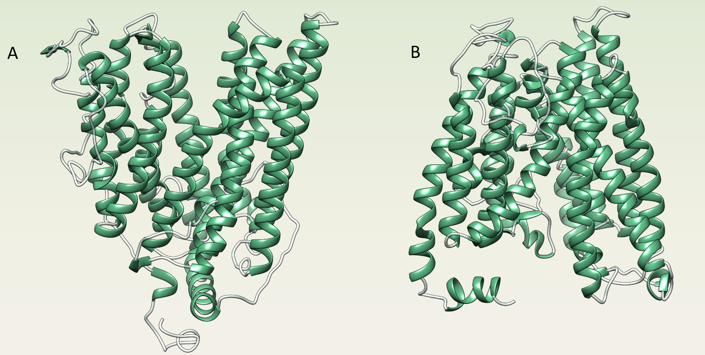
Ferroportin (Fpn) is a membrane protein belonging to the Major Facilitator Superfamily of transporters. It is the only vertebrate iron exporter known so far. Several Fpn mutations lead to the so-called ‘ferroportin disease’ or type 4 haemochromatosis, characterized by two distinct iron accumulation phenotypes depending on whether the mutations affects the protein’s activity or its degradation pathway (1). Despite a general agreement of the scientific community on a 12 transmembrane helices topology, no experimental data are available on human Fpn (HsFpn) three-dimensional structure. Thus, important features of HsFpn remain to be clarified. Recently, the crystal structures of a HsFpn homologue from the predatory Gram-negative bacterium Bdellovibrio bacteriovorus (BbFPN), in both the outward- (Figure 1 A) and inward-open states (Figure 1 B), has been reported (2). The residues essential for iron binding and transport in HsFpn are conserved in BbFPN (3). The conservation of these functionally relevant residues prompted us to exploit the two BbFPN structures to construct reliable models of HsFPN.
Methods
The structural models of HsFpn in were built in both the outward- and inward-open states through the ab initio/threading strategy implemented in the I-TASSER server (4). The overall quality of the models generated has been evaluated using PROCHECK (5) and the model quality parameters provided in the I-TASSER output, such as the C-score. Putative iron binding sites have been detected using LIBRA (6).
Results
The models display the typical fold of MFS proteins with 12 TMs spanning the membrane and the N- and C-termini located on the intracellular side (Figure 1). LIBRA analysis of the models has led to the identification of potential iron binding sites in the inward-open state allowing to propose an iron traslocation mechanism. Further, the outward-open model uncovers details of the interaction site of the peptide hormone hepcidin, a regulator of HsFpn function. Fin ally, the HsFPN models provide a mechanistic interpretation for the disease-related mutations that cause hereditary hemochromatosis.
Author Comment
This is an abstract which has been accepted for the BITS2016 Meeting
Supplemental Information
Figure 1
Schematic representation of the structural models of human Fpn in outwar-open (A) and inward-open states (B).