Endothelial cell-initiated extravasation of cancer cells visualized in zebrafish
- Published
- Accepted
- Subject Areas
- Cell Biology, Oncology
- Keywords
- metastasis, cancer cell, extravasation, zebrafish, endothelial cell, tumor cell, VEGF, sunitinib, fluorescence, in vivo imaging
- Copyright
- © 2014 Kanada et al.
- Licence
- This is an open access article distributed under the terms of the Creative Commons Attribution License, which permits unrestricted use, distribution, reproduction and adaptation in any medium and for any purpose provided that it is properly attributed. For attribution, the original author(s), title, publication source (PeerJ PrePrints) and either DOI or URL of the article must be cited.
- Cite this article
- 2014. Endothelial cell-initiated extravasation of cancer cells visualized in zebrafish. PeerJ PrePrints 2:e500v1 https://doi.org/10.7287/peerj.preprints.500v1
Abstract
The extravasation of cancer cells, a key step for distant metastasis, is thought to be initiated by disruption of the endothelial barrier by malignant cancer cells. An endothelial covering-type extravasation of cancer cells in addition to conventional cancer cell invasion-type extravasation was dynamically visualized in a zebrafish hematogenous metastasis model. The inhibition of VEGF-signaling impaired the invasion-type extravasation via inhibition of cancer cell polarization and motility regulated by an intracellular signaling. Paradoxically, the inhibition of VEGF-signaling showed the promotion, rather than the inhibition, of the endothelial covering-type extravasation of cancer cells, with structural changes in the endothelial walls. These findings may be a clue to the full understanding of the metastatic process as well as the metastatic acceleration by antiangiogenic reagents observed in preclinical studies.
Author Comment
This is a submission to PeerJ for review.
Supplemental Information
Assessing the morphological Recovery of VEGF-depleted Cells by Cancer Cell-cultured Medium
Twenty-four h after the siRNA transfection, culture medium was replaced with fresh medium (left) or cultured medium in which normal RFP-HeLa cells were cultured for 36 h (right), and then siRNA-treated cells were cultured for another 24 h. Phase contrast images show the RFP-HeLa cells in the polymer-bottom dishes 48 h after the siRNA transfection. Bar, 20 μm.
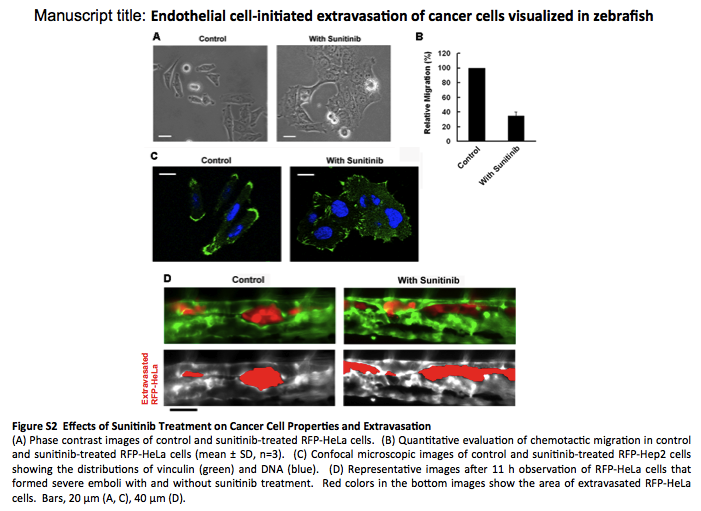
Effects of Sunitinib Treatment on Cancer Cell Properties and Extravasation
(A) Phase contrast images of control and sunitinib-treated RFP-HeLa cells. (B) Quantitative evaluation of chemotactic migration in control and sunitinib-treated RFP-HeLa cells (mean ± SD, n=3). (C) Confocal microscopic images of control and sunitinib-treated RFP-Hep2 cells showing the distributions of vinculin (green) and DNA (blue). (D) Representative images after 11 h observation of RFP-HeLa cells that formed severe emboli with and without sunitinib treatment. Red colors in the bottom images show the area of extravasated RFP-HeLa cells. Bars, 20 μm (A, C), 40 μm (D).
Cancer Cell Invasion-type Extravasation
Invasion-type extravasation of RFP-HeLa cells that formed severe emboli in the caudal artery of the zebrafish larva. Number, elapsed time in minutes.
Endothelial Covering-type Extravasation
Endothelial covering-type extravasation of RFP-HeLa cells that formed severe emboli in the caudal artery of the zebrafish larva. Number, elapsed time in minutes.
3D image of embolous-forming cancer cells and covering endothelial cells
3D image was created with confocal microscopic images (47 slices, step size: 1 mm) taken at 10 hour postadministration.
Control siRNA-treated Cancer Cells in culture
Phase contrast images of RFP-HeLa cells treated with control siRNA in the polymer-bottom dish. Number, elapsed time in minutes.
VEGF-depleted Cancer Cells in culture
Phase contrast images of RFP-HeLa cells treated with siRNA against VEGF in the polymer-bottom dish. Number, elapsed time in minutes.
Extravasation of VEGF-depleted Cancer Cells
Extravasation of the VEGF-depleted RFP-HeLa. Number, elapsed time in minutes.
Extravasation of Sunitinib-treated Cancer Cells
Extravasation of RFP-HeLa cells in the presence of sunitinib. Number, elapsed time in minutes.