A new Python library to analyse skeleton images confirms malaria parasite remodelling of the red blood cell membrane skeleton
- Published
- Accepted
- Subject Areas
- Biochemistry, Bioinformatics, Parasitology, Computational Science
- Keywords
- Python, skeleton analysis, malaria, red blood cell, cytoskeleton
- Copyright
- © 2018 Nunez-Iglesias et al.
- Licence
- This is an open access article distributed under the terms of the Creative Commons Attribution License, which permits unrestricted use, distribution, reproduction and adaptation in any medium and for any purpose provided that it is properly attributed. For attribution, the original author(s), title, publication source (PeerJ Preprints) and either DOI or URL of the article must be cited.
- Cite this article
- 2018. A new Python library to analyse skeleton images confirms malaria parasite remodelling of the red blood cell membrane skeleton. PeerJ Preprints 6:e3521v1 https://doi.org/10.7287/peerj.preprints.3521v1
Abstract
We present Skan (Skeleton analysis), a Python library for the analysis of the skeleton structures of objects. It was inspired by the “analyse skeletons” plugin for the Fiji image analysis software, but its extensive Application Programming Interface (API) allows users to examine and manipulate any intermediate data structures produced during the analysis. Further, its use of common Python data structures such as SciPy sparse matrices and pandas data frames opens the results to analysis within the extensive ecosystem of scientific libraries available in Python. We demonstrate the validity of Skan’s measurements by comparing its output to the established Analyze Skeletons Fiji plugin, and, with a new scanning electron microscopy (SEM)-based method, we confirm that the malaria parasite Plasmodium falciparum remodels the host red blood cell cytoskeleton, increasing the average distance between spectrin-actin junctions.
Author Comment
This is a submission to PeerJ for review.
Supplemental Information
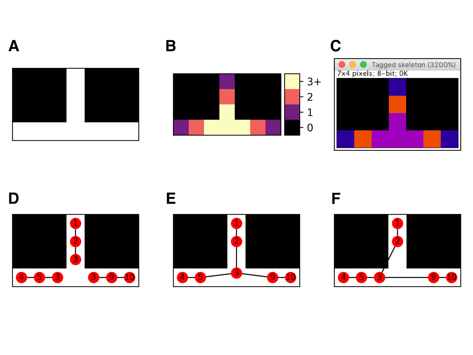
Strategies for resolving skeleton junctions
(A) A minimal skeleton. (B) Skan’s classification of pixels into endpoints, paths, and junctions based on the number of neighbours (1, 2, and 3 or more, respectively). (C) Identical classification in Fiji’s Analyze Skeletons. (D) Skeleton measurement when junctions are assigned an implicit “extent”. (E) Skeleton measurement when all adjacent junction pixels are replaced by their centroid (our default strategy). (F) Skeleton measurement used in Fiji’s Analyze skeletons (mid-2017 version).