Anatomical and biomechanical traits of broiler chickens across ontogeny. 1. Anatomy of the musculoskeletal respiratory apparatus and changes in organ size.
- Published
- Accepted
- Subject Areas
- Agricultural Science, Developmental Biology, Evolutionary Studies, Zoology, Anatomy and Physiology
- Keywords
- broiler, development, breathing, organ, pathology, scaling
- Copyright
- © 2014 Tickle et al.
- Licence
- This is an open access article distributed under the terms of the Creative Commons Attribution License, which permits unrestricted use, distribution, reproduction and adaptation in any medium and for any purpose provided that it is properly attributed. For attribution, the original author(s), title, publication source (PeerJ PrePrints) and either DOI or URL of the article must be cited.
- Cite this article
- 2014. Anatomical and biomechanical traits of broiler chickens across ontogeny. 1. Anatomy of the musculoskeletal respiratory apparatus and changes in organ size. PeerJ PrePrints 2:e342v1 https://doi.org/10.7287/peerj.preprints.342v1
Abstract
Genetic selection for improved meat yields, digestive efficiency and growth rates have transformed the biology of broiler chickens. Modern birds undergo a 50-fold multiplication in body mass in just six weeks, from hatching to slaughter weight. However, this selection for rapid growth and improvements in broiler productivity is also widely thought to be associated with increased welfare problems as many birds suffer from leg, circulatory and respiratory diseases. To understand growth-related changes in musculoskeletal and organ morphology and respiratory skeletal development over the standard six-week rearing period, we present data from post-hatch cadaveric commercial broiler chickens aged 0, 2, 4 and 6 weeks. The heart, lungs and intestines decreased in size for hatch to slaughter weight when considered as a proportion of body mass. Proportional liver size increased in the two weeks after hatch but decreased between 2 and 6 weeks. Breast muscle mass on the other hand displayed strong positive allometry, increasing in mass faster than the increase in body mass. Contrastingly, less rapid isometric growth was found in the external oblique muscle, a major respiratory muscle that moves the sternum dorsally during expiration. Considered together with the relatively slow ossification of elements of the respiratory skeleton, it seems that rapid growth of the breast muscles might compromise the efficacy of the respiratory apparatus. Furthermore, the relative reduction in size of the major organs indicates that selective breeding in meat-producing birds has unintended consequences that may bias these birds toward compromised welfare and could limit further improvements in meat-production and feed efficiency.
Supplemental Information
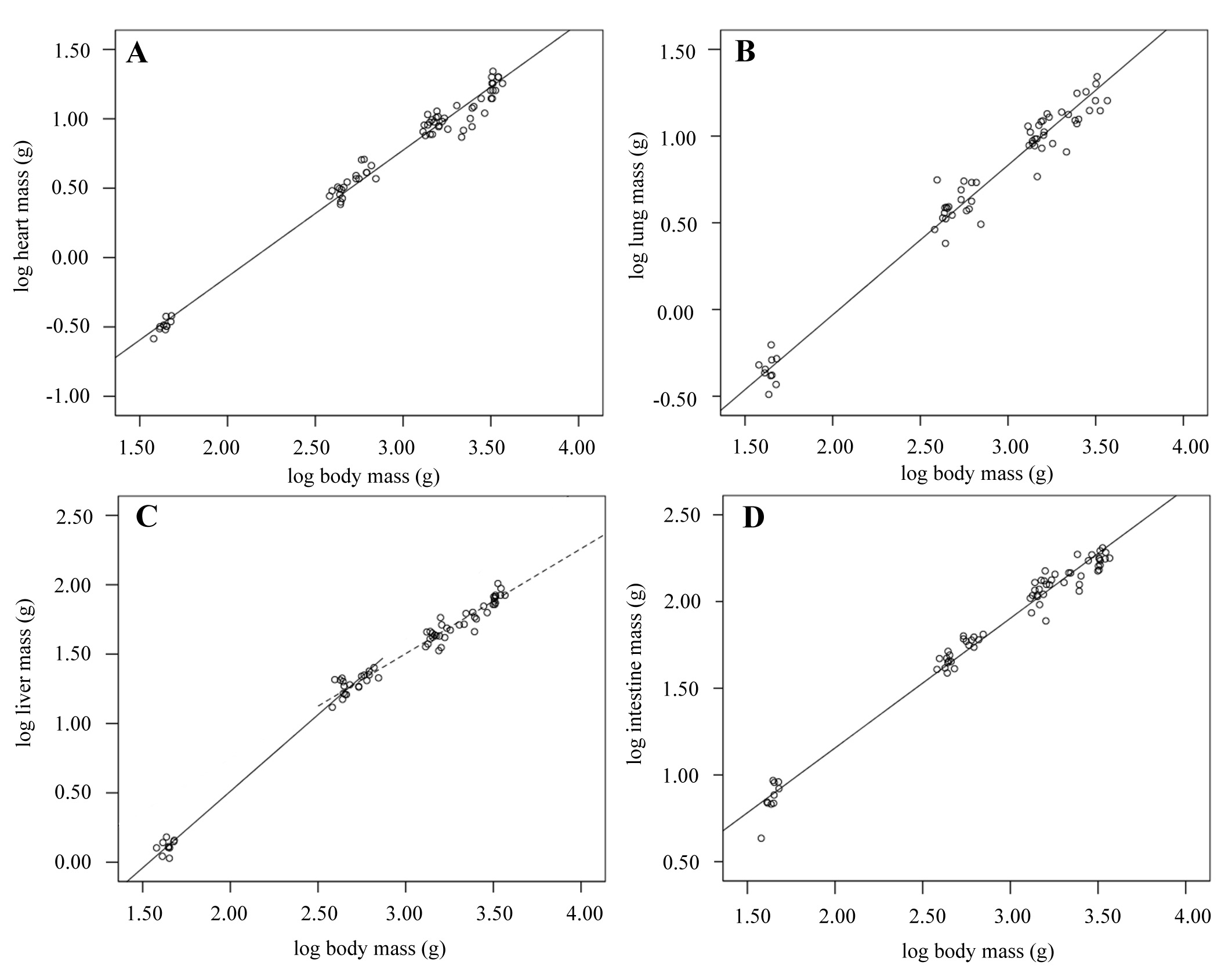
Organ growth during development
Scatter plots showing log transformed organ masses against log body mass over development from hatch to 6-weeks old. The equations describing lines of best fit were: (A) heart: Mb0.91 – 1.96; (n = 69; r2 = 0.98; p < 0.001); (B) lung: Mb0.86 – 1.76 (n = 62; r2 = 0.97; p < 0.001); (C) liver: 0 – 2 weeks old (solid line): Mb1.10 – 1.70 (n = 30; r2 = 0.99; p < 0.001); 2 – 6 weeks old (dashed line): Mb0.76 – 0.77 (n = 59; r2 = 0.95; p < 0.001); (D) intestine: Mb0.75 – 0.34 (n= 69; r2 = 0.98; p < 0.001).
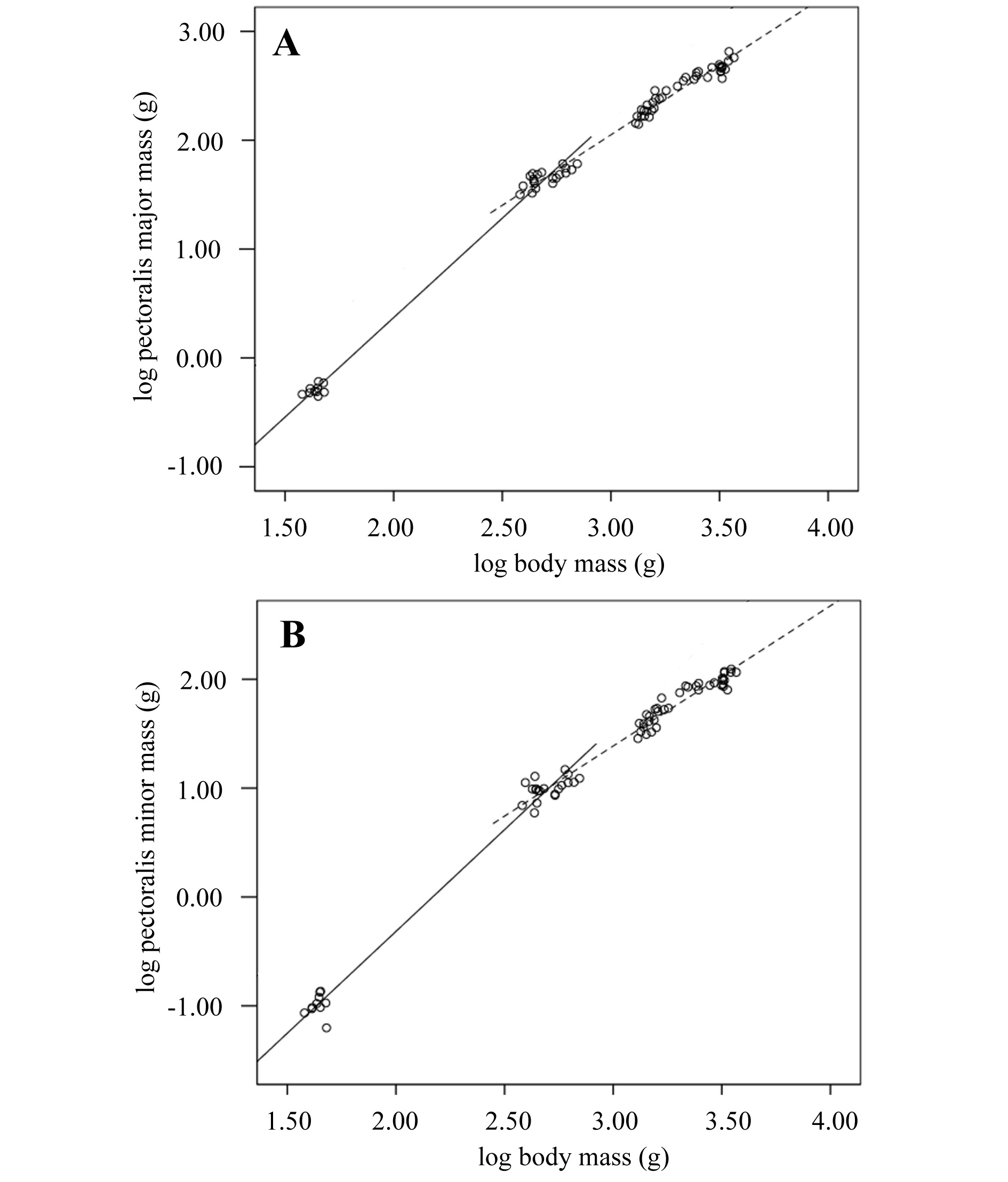
Breast muscle growth during development
Scatter plots showing log transformed pectoralis major and minor masses against log body mass over development from 0 - 2-weeks (solid line) and 2 – 6-weeks old (dashed line). The equations describing lines of best fit were: (A) pectoralis major 0 – 2 weeks: Mb1.83 – 3.28 (n = 30, r2 = 0.99; p < 0.001); 2 – 6 weeks: Mb1.29 – 1.83 (n = 59; r2 = 0.98; p < 0.001).
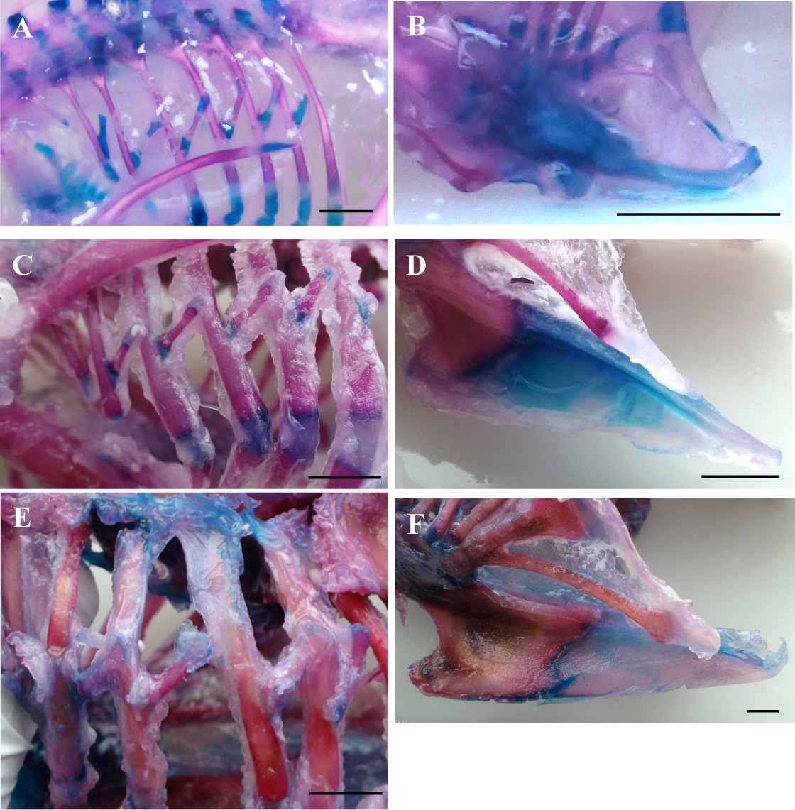
Ossification of the thoracic skeleton
Representative stained skeletons showing the progression of ossification in the vertebral ribs, uncinate processes and sternum. Blue areas correspond to cartilage and red areas to bone. Ossification of ribs and uncinate processes are shown for the hatchling (A and B), 2-week old (C and D) and 6-week old (E and F) birds panels. Ossification of the uncinate processes and sternum remain incomplete at slaughter age. Scale bars represent 10mm.