The effects of Tibetan medicine Zuotai and β-HgS on cytotoxicity and endoplasmic reticular stress-related genes expressions differentiate from HgCl2 in PC-12 cells
- Published
- Accepted
- Subject Areas
- Toxicology, Pharmacology
- Keywords
- cytotoxicity, caspase family genes, endoplasmic reticular stress, mercuric compounds, Tibetan medicine Zuotai
- Copyright
- © 2017 Geng et al.
- Licence
- This is an open access article distributed under the terms of the Creative Commons Attribution License, which permits unrestricted use, distribution, reproduction and adaptation in any medium and for any purpose provided that it is properly attributed. For attribution, the original author(s), title, publication source (PeerJ Preprints) and either DOI or URL of the article must be cited.
- Cite this article
- 2017. The effects of Tibetan medicine Zuotai and β-HgS on cytotoxicity and endoplasmic reticular stress-related genes expressions differentiate from HgCl2 in PC-12 cells. PeerJ Preprints 5:e3308v1 https://doi.org/10.7287/peerj.preprints.3308v1
Abstract
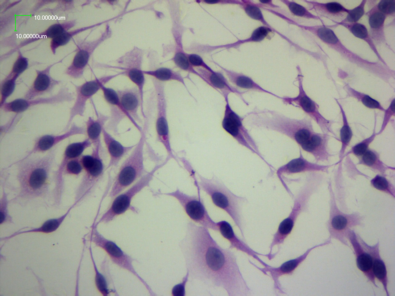
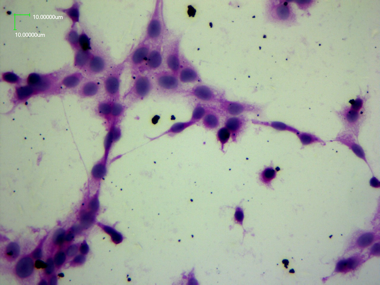
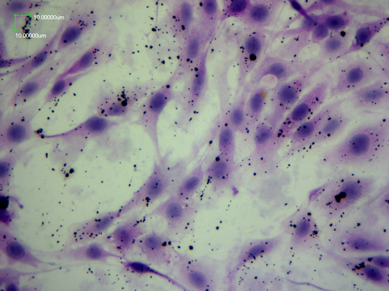
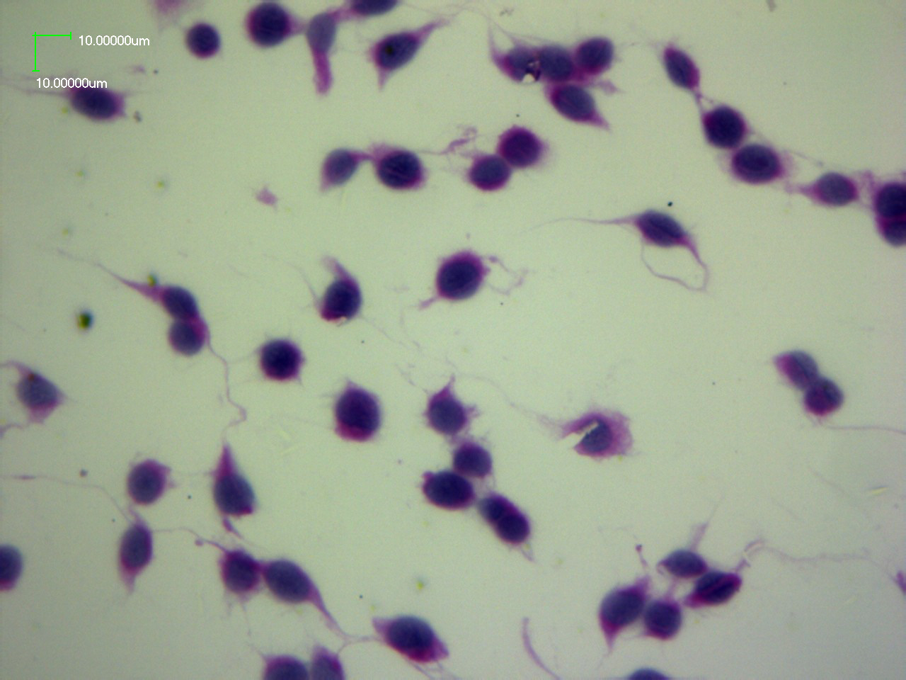
Backgroud. Mercuric sulfide (HgS) has been used in traditional medicines for thousands of years, such as cinnabar, Zuotai and so on. Increasing research supports that HgS is different from mercury chloride (HgCl2) and methylmercury (MeHg) in toxicity and pharmacology, due to the differences in chemical forms. However, the differences of the cytotoxicity and endoplasmic reticulum stress (ERs) among Zuotai, β-HgS and HgCl2 is still unclear. Methods. This study was designed to investigate the differential effects of Zuotai (0.250 g/L, 0.116 g Hg/L), β-HgS (0.135 g/L, 0.116 g Hg/L) and HgCl2 (0.0160 g/L, 0.0116 g Hg/L) on cell cytotoxicity and ERs. In this study, Cell viability, cellular morphology and the expressions of caspase family genes, ERs-related genes and metal sensitive genes were examined in PC-12 cells exposed with or without drugs. Results. Cell viability in Zuotai and β-HgS groups were higher than HgCl2 group (p<0.05), and the cell viability were 94.79 ± 1.958%, 99.27 ± 1.328% and 65.82 ± 3.415% in Zuotai, β-HgS and HgCl2 groups, respectively. Zuotai reduced the adhesion of cells slightly, β-HgS did not change cellular morphology, and HgCl2 produced cell shrinkage and rounded up. Thus, Zuotai and β-HgS produced less cytotoxicity than HgCl2. To examine the signaling pathways activated by Zuotai, β-HgS and HgCl2, caspase family genes, ERs-related genes and metal sensitive genes expressions were examined. Zuotai decreased the expressions of Cas3, Cas8 and Cas9 and upregulated the expression of Cas12, while β-HgS decreased the caspase family genes (Cas3, Cas8, Cas9 and Cas12) expressions. Meanwhile, all three drugs upregulated the expressions of ERs-related genes (GRP78, Gadd45a and Gadd45b) and metal sensitive genes (MT-1 and HO-1). Conclusion. Zuotai and β-HgS had less cytotoxicity than HgCl2. The cell viability of Zuotai and β-HgS were significantly higher than HgCl2, and Zuotai and β-HgS inhibited caspase activity while HgCl2 produced cell death through the caspase independent pathway. Zuotai, β-HgS and HgCl2 triggered ERs and metal sensitive genes expressions. Taken together, Zuotai and β-HgS were different from HgCl2 in cellular toxicity, and the effects of Zuotai and β-HgS at a high dose were both protective and destructive.
Author Comment
This is a submission to PeerJ for review.