The first juvenile specimens of Plateosaurus engelhardti from Frick, Switzerland: Isolated neural arches and their implications for developmental plasticity in a basal sauropodomorph
- Published
- Accepted
- Subject Areas
- Paleontology
- Keywords
- Late Triassic, Norian, Switzerland, Basal Sauropodomorpha, Plateosaurus engelhardti, juvenile, neurocentral suture closure, bone histology
- Copyright
- © 2014 Hofmann et al.
- Licence
- This is an open access article distributed under the terms of the Creative Commons Attribution License, which permits unrestricted use, distribution, reproduction and adaptation in any medium and for any purpose provided that it is properly attributed. For attribution, the original author(s), title, publication source (PeerJ PrePrints) and either DOI or URL of the article must be cited.
- Cite this article
- 2014. The first juvenile specimens of Plateosaurus engelhardti from Frick, Switzerland: Isolated neural arches and their implications for developmental plasticity in a basal sauropodomorph. PeerJ PrePrints 2:e325v1 https://doi.org/10.7287/peerj.preprints.325v1
Abstract
The dinosaur Plateosaurus engelhardti is the most abundant dinosaur in the Late Triassic of Europe and the best known basal sauropodomorph. Plateosaurus engelhardti was one of the first sauropdomorph dinosaurs to display a large body size. Remains can be found in the Norian stage of the Late Triassic in over 40 localities in Central Europe (France, Germany, Greenland and Switzerland). Since the first discovery of P. engelhardti no juvenile specimens of this species had been found. Here we describe the first remains of juvenile individuals, isolated cervical and dorsal neural arches. These were separated postmortem from their respective centra because of unfused neurocentral sutures. However the specimens share the same neural arch morphology found in adults. Morphometric analysis suggests a body lengths of the juvenile indivduals that is greater than those of most adult specimens. This supports the hypothesis of developmental plasticity in Plateosaurus engelhardti that previously had been based on histological data only. Alternative hypotheses for explaining the poor correlation between ontogenetic stage and size in this taxon are multiple species or sexual morphs with little morphological variance or time-averaging of individuals from populations differing in body size.
Supplemental Information
Table S1: Zygapophyses lengths of the juvenile specimens MSF 11.3., MSF 5B, MSF 23 and SMNS 13200.
Zygapophyses lengths in mm of the juvenile specimens MSF 11.3., MSF 5B, MSF 23 and SMNS 13200 with their respective position in the vertebral column. As noted in the description some positions can be recognized twice. The zygapophyses lengths in parentheses belong to the longer neural arch of those to be found twice in the sample. The specimen numbers of the 11.3. specimens is listed in Table 1. D3 (MSF 11.3.376/77.1 mm and MSF 11.3.360/86.7 mm). D5 (MSF 11.3.167/94.2 mm and MSF 11.3.067/101.1 mm. D6 (MSF 11.3.095/108.5 mm and MSF 11.3.107/109.2 mm). D10/D11 (MSF 11.3.241/110.8 mm and MSF 11.3.303/121.6 mm). Gaps are left, where data is missing due to preservation or the accessibility is was not given.
Table S2: Zyg/Fe ratios of MSF 5B, MSF 23 and SMNS 13200.
Table S2 shows the calculated ratio in percent between the zygapophyses length and the femur length of the referred specimens. The ratios within on specimen and compared to each other specimen follow a determined scheme of zygapohyseal length explaining the wide range between 12.5 – 28.3 %. With the help of the ratios we were able to define a new proxy for the determination of a range of femur lengths for the juvenile MSF 11.3. specimens.
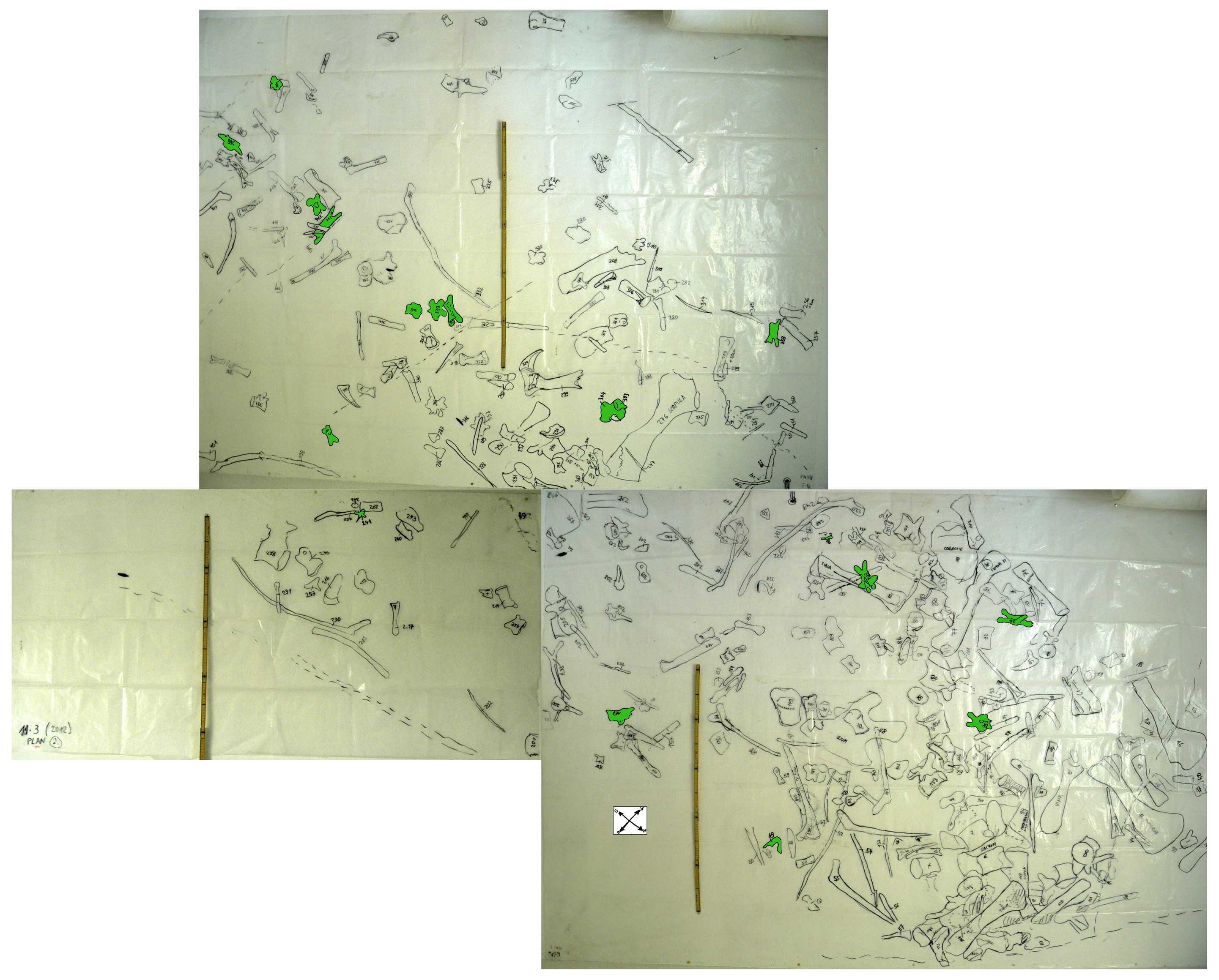
Figure S1: Foil plan of bone field 11.3.
The foil plan shows the isolated neural arches found in bone field 11.3. The neural arches are colored in green. The plan clearly shows that the neural arches were distributed over the whole area with no recognizable connection to each other and no centra lying next to them. The yardstick measures 1m.
Figure S2: Specimen MSF 11.3.317 (axis) and MSF 11.3.258 (C3)
Plateosaurus engelhardti from Frick, Switzerland. Anterior neural arches of late juveniles. A-C: MSF 11.3.317 (axis) in A, left lateral view; B, dorsal view and C, ventral view. Specimen MSF 11.3.317 shows prezygapophyses facets being smaller and shorter than those of the postzygapophyses. The spof is the only fossa developed. D-F: MSF 11.3.258 (C3) in D, dorsal view; E, ventral view and F, right lateral view. The spof in MSF 11.3.258 gets deeper and the sprf developed. Note the zipper-like structures on the pedicels in ventral view. Furthermore the specimen shows dessication cracks in dorsal view allover on the neural arch. See text for abbrevations. Scale bars measure 1 cm.
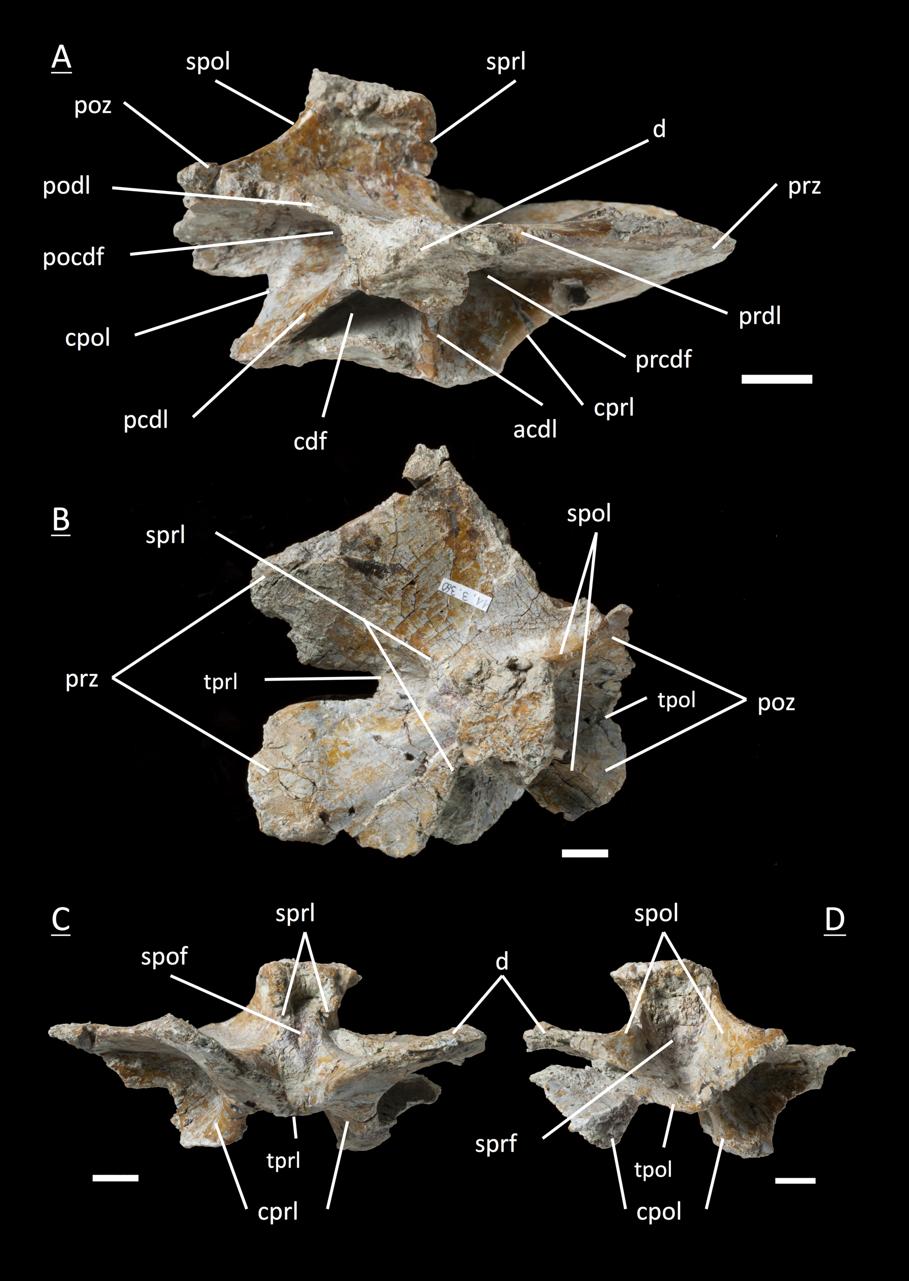
Figure S3: Specimen MSF 11.3.371 (C4)
Plateosaurus engelhardti from Frick, Switzerland. Anterior cervical neural arch of a late juvenile. A-E: MSF 11.3.371 (C4) in A, left lateral view; B, dorsal view; C, ventral view; D, anterior view and E, posterior view. A partly preserved diapophysis on the right lateral side is visible. See text for abbreviations. Scale bars measure 1 cm.
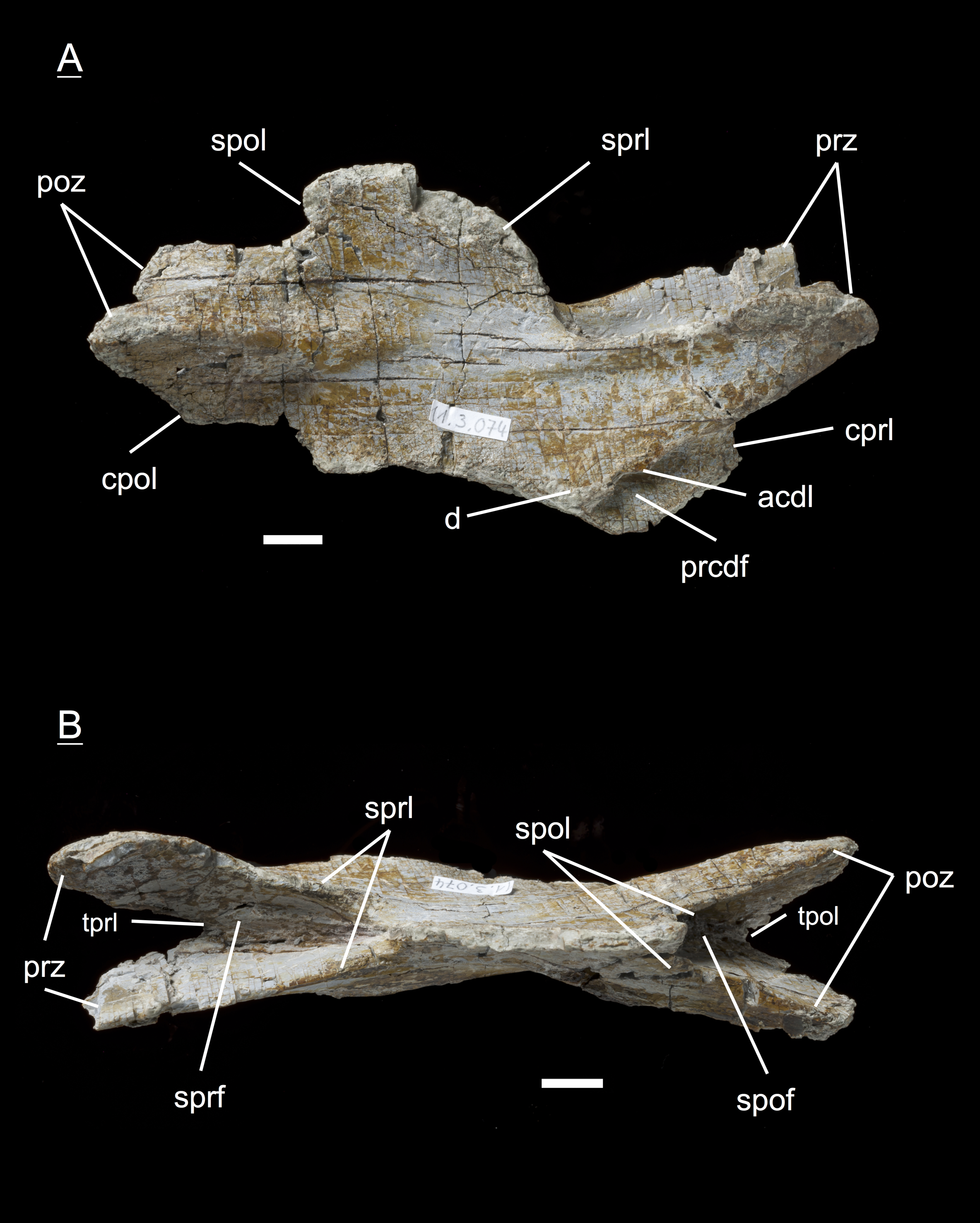
Figure S4: Specimen MSF 11.3.074 (C6)
Plateosaurus engelhardti from Frick, Switzerland. Posterior cervical neural arch of a late juvenile. A-B: MSF 11.3.074 (C6) in A, right lateral view and D, dorsal view. Articular facets of the prezygapophyses and postzygapophyses are rough, suggesting a cover by cartilage. The diapophyses is well developed. Specimen MSF 11.3.074 shows dessication cracks in right lateral view allover on the neural arch. See text for abbreviations. Scale bars measure 1 cm.
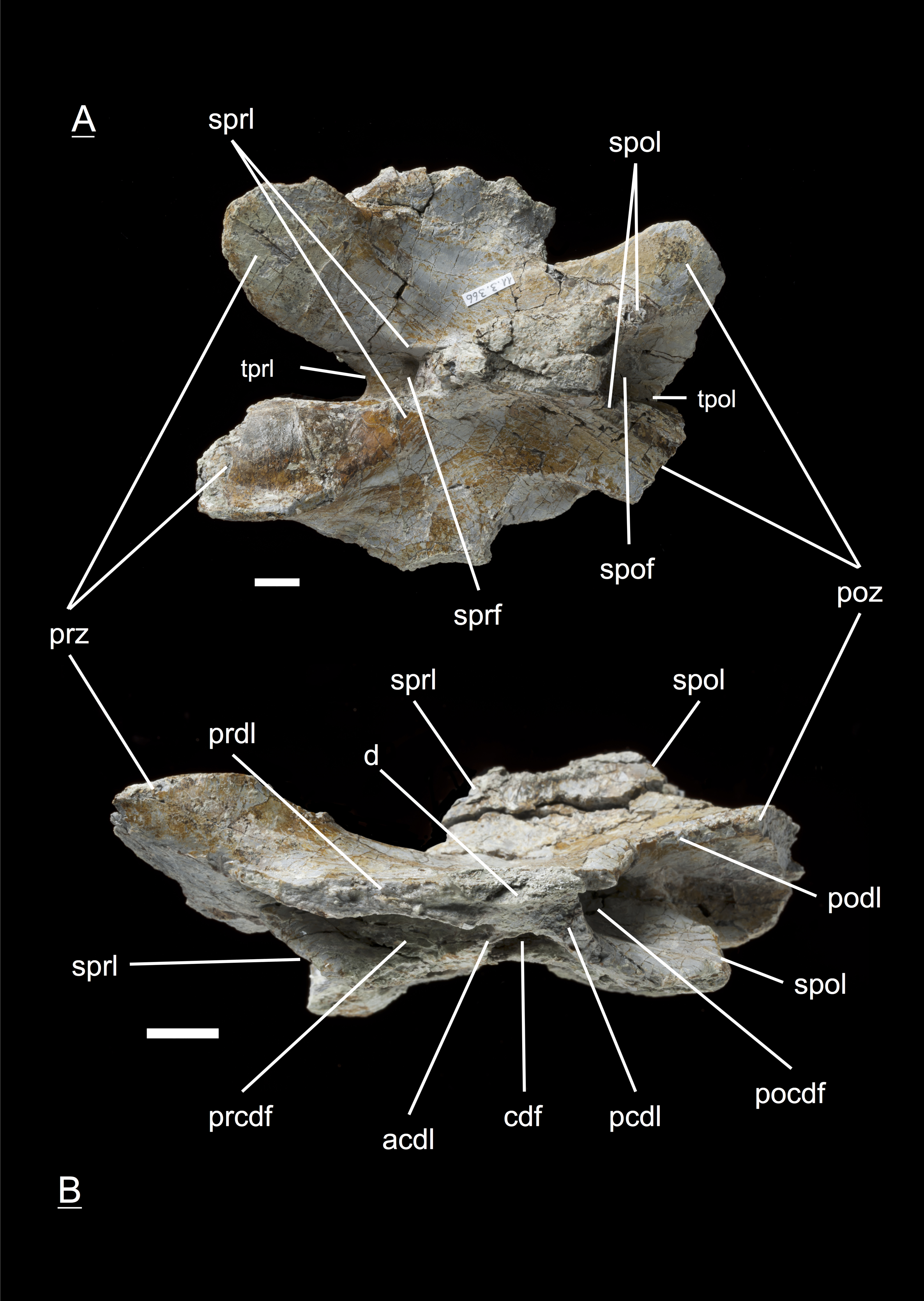
Figure S5: Specimen MSF 11.3.366 (C10)
Plateosaurus engelhardti from Frick, Switzerland. Posterior cervical neural arch of a late juvenile. A-B: MSF 11.3.366 (C10) in A, dorsal view and B, left lateral view. Specimen MSF 11.3.366 represents the cervicodorsal transition of posteriormost cervicals and anteriormost dorsals very well. Transverse processes (diapophyses and parapophyses) are changing in shape, size and function. Therefore all of the diapophyseal laminae and fossae are well developed. Dessication cracks are present in left lateral view on the prz and poz on both sides of the neural arch. See text for abbreviations. Scale bars measures 1 cm.
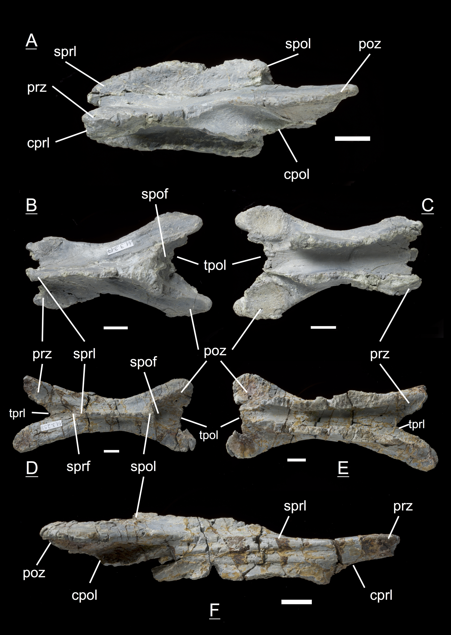
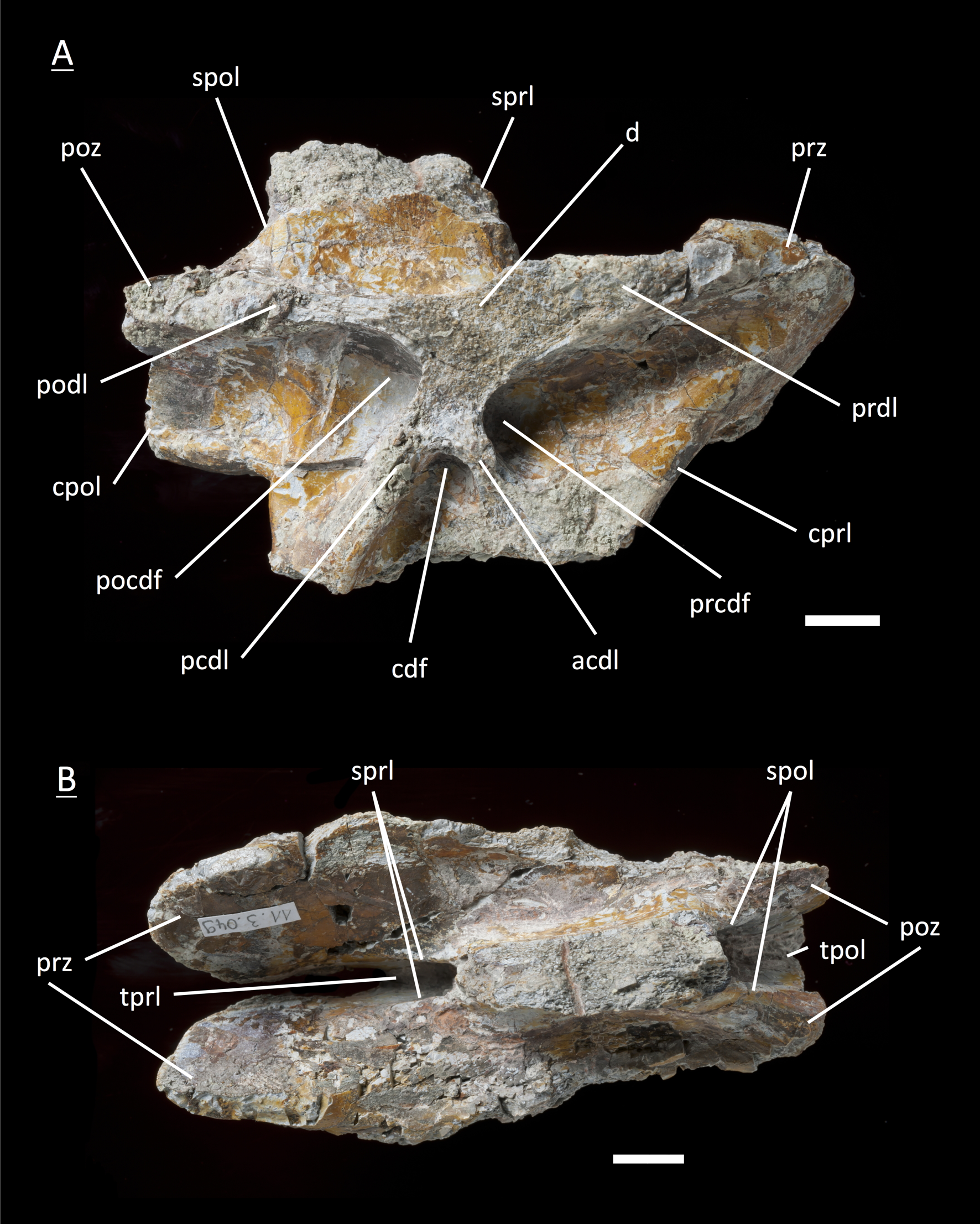
Figure S6: Specimen MSF 11.3.360 (D3)
Plateosaurus engelhardti from Frick, Switzerland. Anterior dorsal neural arch of late a juvenile. A-D: MSF 11.3.360 (D3) in A, right lateral view; B, dorsal view; C, anterior view and D, posterior view. Specimen MSF 11.3.360 has the shortest and thickest neural spine of all neural arches studied. Parapophysis articular facets slightly become visible. See text for abbreviations. Scale bars measure 1 cm.
Figure S7: Specimen MSF 11.3.376 (D3)
Plateosaurus engelhardti from Frick, Switzerland. Anterior dorsal neural arch of a late juvenile. A-C: MSF 11.3.376 (D3) in A, right lateral view; B, dorsal view and C, posterior view. See text for abbreviations. This specimen shows the same diagnostic characters as specimen MSF 11.3.360 except for very well developed parapophysis articular facets. It is unusual since these facets first show up in the fifth or sixth dorsal. Scale bars measure 1 cm.
Figure S8: Specimen MSF 11.3.049 (D4)
Plateosaurus engelhardti from Frick, Switzerland. Anterior dorsal neural arch of a late juvenile. MSF 11.3.049 (D4) in A, right lateral view and B, dorsal view. No parapophyses are visible. All of the diapophseal laminaw and fossae ware well developed. See text for abbreviations. Scale bars measure 1 cm.
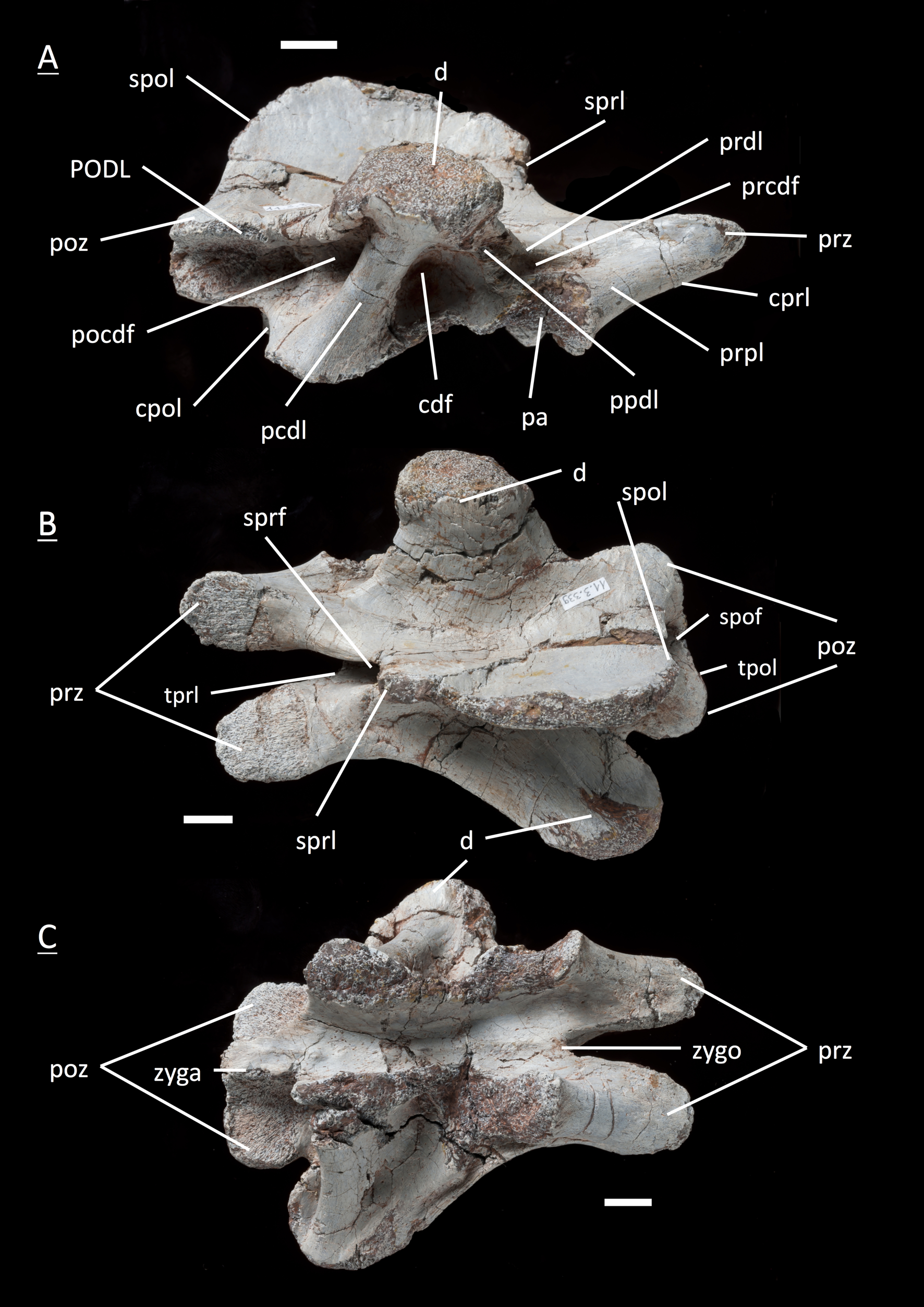
Figure S9: Specimen MSF 11.3.067 (D5)
Plateosaurus engelhardti from Frick, Switzerland. Anterior dorsal neural arch of a late juvenile. MSF 11.3.067 (D5) in A, left lateral view; B, dorsal view and C, ventral view. All laminae and fossae are still well developed, but the prcdf begins to decrease in size and extent due to the parapophysis articular facets moving upwards onto the neural. Specimen MSF 11.3.067 shows dessication cracks in dorsal view on the left lateral prz and on the right lateral d. See text for abbreviations. Scale bars measure 1 cm.
Figure S10: Specimen MSF 11.3.167 (D5)
Plateosaurus engelhardti from Frick, Switzerland. Anterior dorsal neural arch of a late juvenile. MSF 11.3.167 (D5) in A, right lateral view; B, dorsal view and C, ventral view. On the neural arch of this specimen the parapophysis articular facet is well visible. The cdf contains crushed bone caused by crushing. Dessication cracks are present in dorsal view on the right lateral prezygapophyis and in ventral view on the pedicels on both sides. See text for abbreviations. Scale bars measure 1 cm.
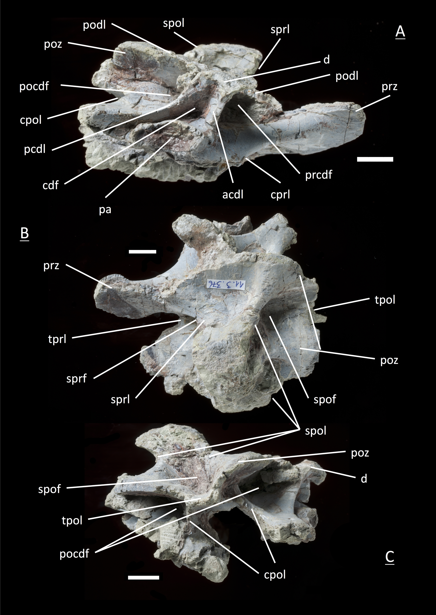
Figure S11: Specimen MSF 11.3.095 (D6)
Plateosaurus engelhardti from Frick, Switzerland. Middle dorsal neural arch of a late juvenile. MSF 11.3.095 (D6) in A, left lateral view; B, dorsal view and C, ventral view. The parapohysis articular facet displaces the acdl, giving rise to the ppdl, the acpl and prpl. Due to this change of laminae the prcdf becomes smaller in extent. Specimen MSF 11.3.095 shows dessication cracks in left lateral view, dorsal view as well as ventral view. See text for abbreviations. Scale bars measure 1 cm.
Figure S12: Specimen MSF 11.3.107 (D6)
Plateosaurus engelhardti from Frick, Switzerland. Middle dorsal neural arch of a late juvenile. MSF 11.3.107 (D6) in A, right lateral view; B, ventral view and C, posterior view. All of the characters found coincide with those of MSF 11.3.095. See text for abbreviations. Scale bars measure 1 cm.
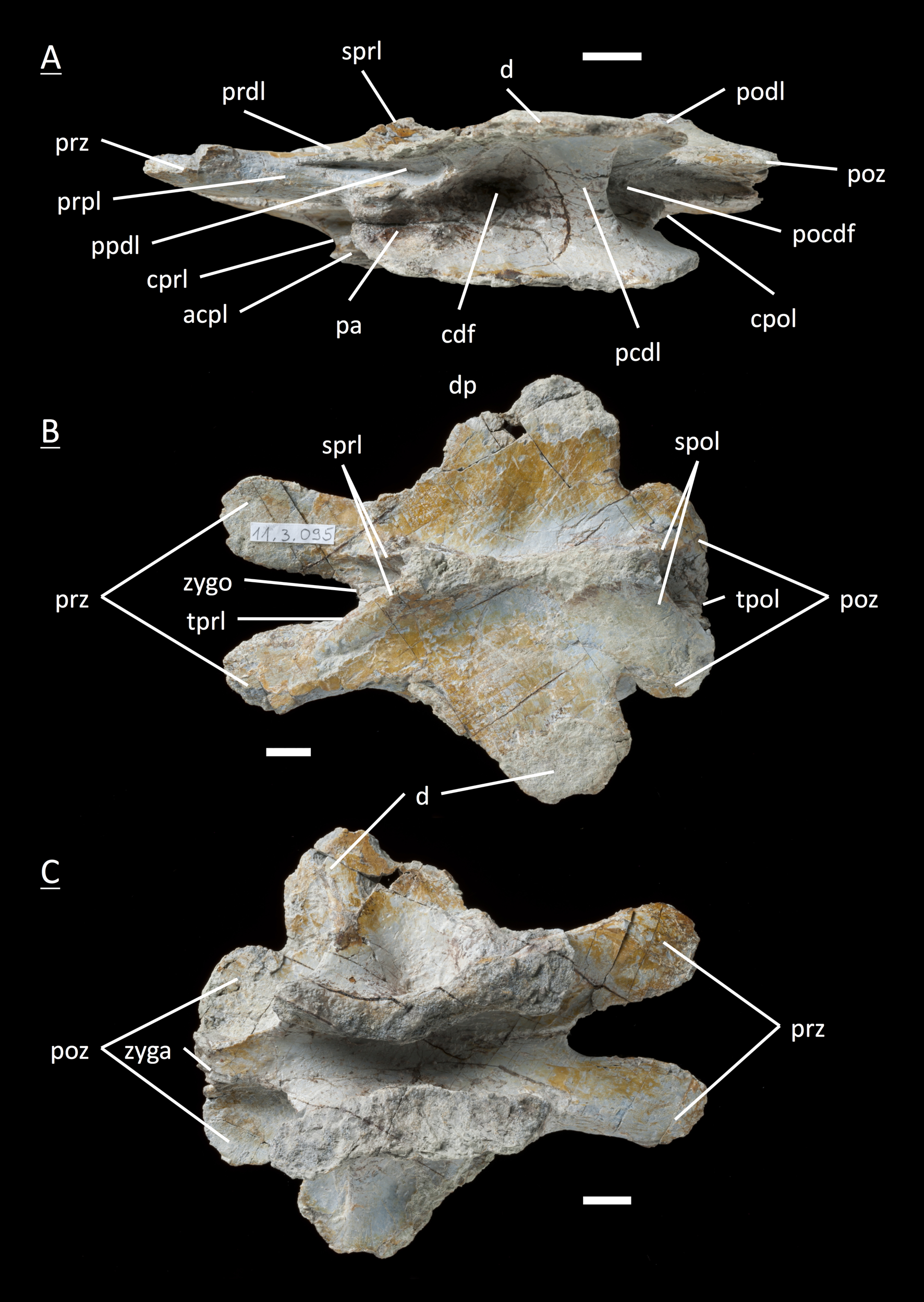
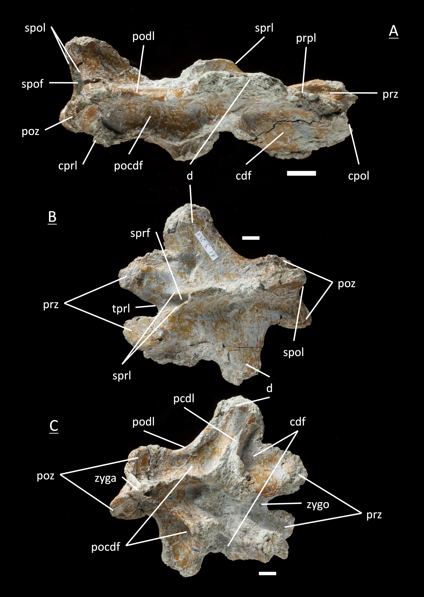
Figure S13: Specimen MSF 11.3.339 (D7)
Plateosaurus engelhardti from Frick, Switzerland. Middle dorsal neural arch of a late juvenile. MSF 11.3.339 (D7) in A, right lateral view; B, dorsal view and C, ventral view. The prcdf is extremely diminished in comparison to anterior dorsal neural arches. The articular surfaces of the prezygapophyses, postzygapophyses, and diapophyses display very rough articular surfaces once being covered by cartilage. The abrasion is an indicator of osteological immaturity. Specimen MSF 11.3.339 shows dessication cracks in ventral view on the left lateral prz. See text for abbreviations. Scale bars measure 1 cm.
Figure S14: Specimen MSF 11.3.241 (D10/D11)
Plateosaurus engelhardti from Frick, Switzerland. Middle/Posterior dorsal neural arch of a late juvenile. MSF 11.3.241 (D10/D11) in A, right lateral view; B, dorsal view and C, ventral view. The diapophyses are broad and extensive. Only two diapophyseal fossae are present (cdf and pocdf). Zygosphene and zygantrum are very distinctive. See text for abbreviations. Scale bars measure 1 cm.
Figure S15: Specimen MSF 11.3.303 (D10/D11)
Plateosaurus engelhardti from Frick, Switzerland. Middle/Posterior dorsal neural arch of a late juvenile. MSF 11.3.303 (D10/D11) in A, right lateral view; B, dorsal view and C, ventral view. In ventral view a partly preserved posterior caudal vertebrae (MSF 11.3.304) is cemented to specimen MSF 11.3.303. See text for abbreviations. Scale bars measure1 cm.