Dose dependent role of Emodin and BTB14431 in suspension colon cancer model in rats
- Published
- Accepted
- Subject Areas
- Drugs and Devices, Oncology, Surgery and Surgical Specialties
- Keywords
- tumor, Angiogenesis, BTB14431, Emodin, rats
- Copyright
- © 2014 Rogalla et al.
- Licence
- This is an open access article distributed under the terms of the Creative Commons Attribution License, which permits unrestricted use, distribution, reproduction and adaptation in any medium and for any purpose provided that it is properly attributed. For attribution, the original author(s), title, publication source (PeerJ PrePrints) and either DOI or URL of the article must be cited.
- Cite this article
- 2014. Dose dependent role of Emodin and BTB14431 in suspension colon cancer model in rats. PeerJ PrePrints 2:e304v1 https://doi.org/10.7287/peerj.preprints.304v1
Abstract
Background. An “In Silico 2D/3D Conformer Screening” for structural similar antitumor substances to Curcumin was carried out and the novel antrachinone BTB14431 was found. Emodin, contained in several Chinese medical plants and BTB14431 are known to be potential inhibitors of the COP9-signalosome - stabilizing the tumor suppressor protein p53. The aim of this study was to analyze the suppressing effects on colorectal cancer in a standardized rat model (WAG/Rij).
Methods. A suspension of CC531 colon cancer cells was applied to the cecum after laparotomy and, additionally, at the back of animals. Therapy was conducted twice daily for 7 days, with increasing doses of BTB14431, Emodin and with isotone sodium chloride solution (control) intravenously (iv) or intraperitoneally (ip). Therapy was initiated the day of tumor cell application. Peripheral blood samples were taken before surgery and on day 7. 21 days after the end of therapy, the animals were euthanized and tumor growth was evaluated.
Results. Data showed a downward trend of the total tumor growth after iv and ip treatment with low doses of BTB14431 and Emodin. Differential blood analysis showed apoptosis, but no major changes in hemogram. Increasing doses of Emodin elevated total mortality rate exponentially.
Conclusions.Although apoptosis was verified, no significant tumor suppressing effects could be observed for iv and ip treatment of both agents in our model. This stays in contrast to former in vitro studies. Agents remain viable novel substances. They will be the subject of upcoming studies. Additional data is needed to evaluate the significance of the “In Silico Screening” to identify potential in vivo anti-tumor drugs.
Author Comment
This document will be submitted to PeerJ for review. The data are unpublished so far.
Supplemental Information
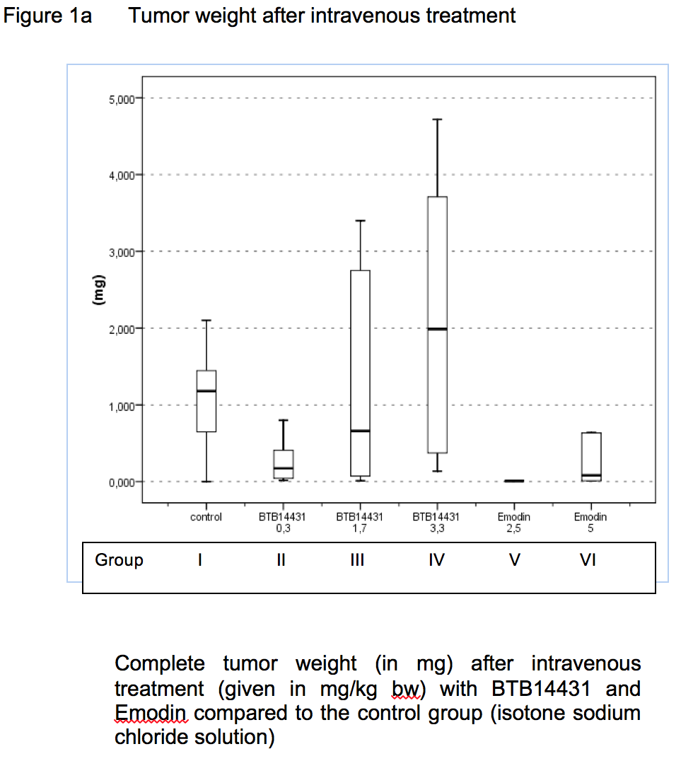
Figure 1a. Tumor weight after intravenous treatment
Complete tumor weight (in mg) after intravenous treatment (given in mg/kg bw) with BTB14431 and Emodin compared to the control group (isotone sodium chloride solution)
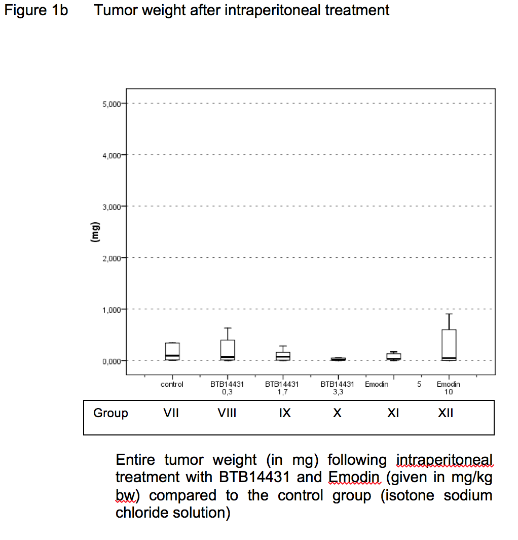
Figure 1b. Tumor weight after intraperitoneal treatment
Entire tumor weight (in mg) following intraperitoneal treatment with BTB14431 and Emodin (given in mg/kg bw) compared to the control group (isotone sodium chloride solution)
Figure 2a. Hemogram after intravenous therapy.
For identification of the groups see table 1 (Roman numerals, e.g. group one left two bars). Figure shows relative rate of leukocyte count (mean) in percent of 100%: pre op (0, left bar) and after last treatment (e.g. second bar). Neutrophil count increases whereas monocyte and lymphocyte count decreases during treatment. Eosinophil count increases in a lower rate.
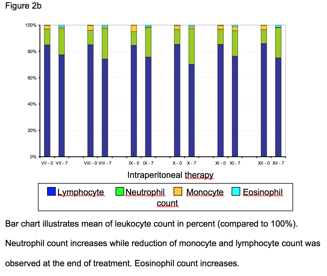
Figure 2b. Hemogram after intraperitoneal therapy.
Bar chart illustrates mean of leukocyte count in percent (compared to 100%). Neutrophil count increases while reduction of monocyte and lymphocyte count was observed at the end of treatment. Eosinophil count increases.
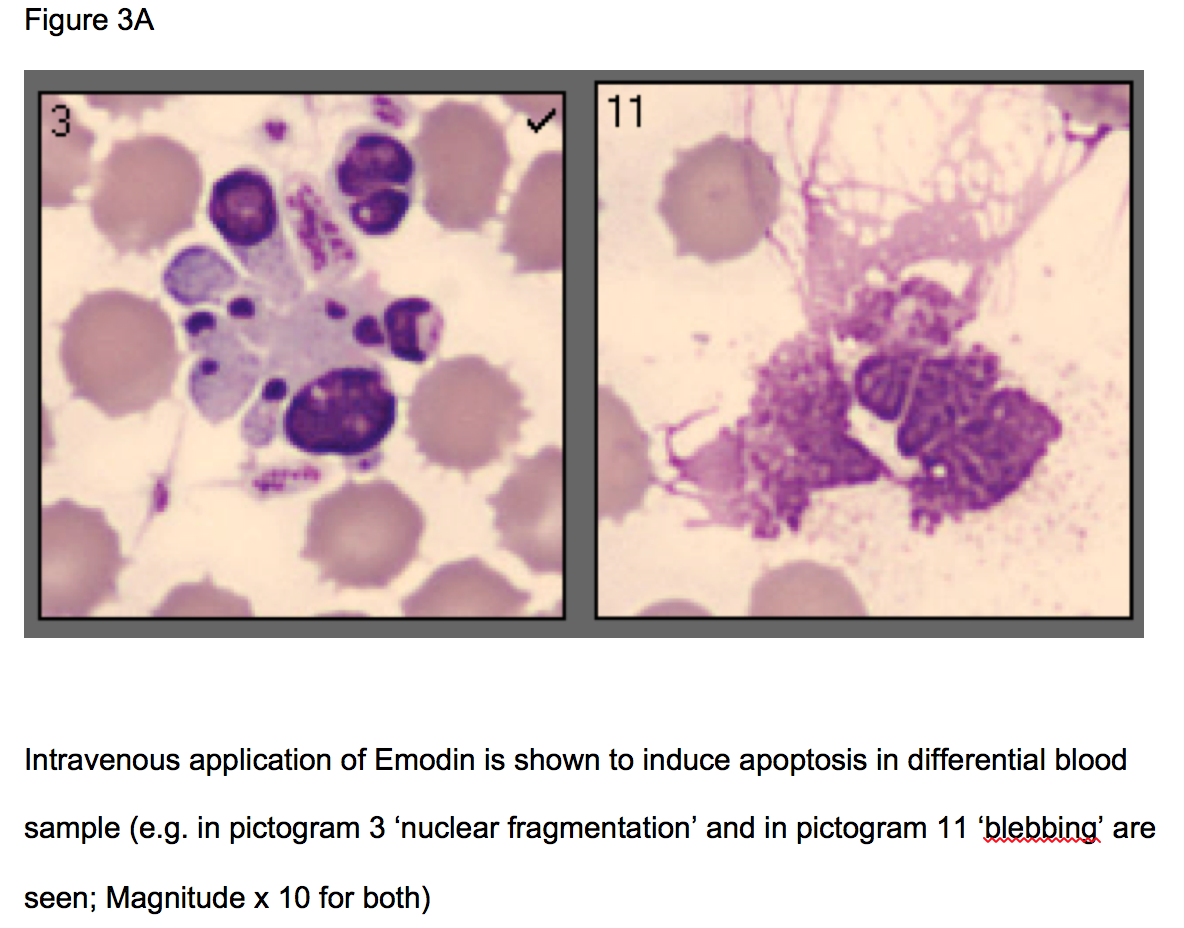
Figure 3a. Apoptosis on granulocytes caused by Emodin
Intravenous application of Emodin is shown to induce apoptosis in differential blood sample (e.g. in pictogram 3 ‘nuclear fragmentation’ and in pictogram 11 ‘blebbing’ are seen; Magnitude x 10 for both)
Figure 3b. Apoptotic effects on granulocytes by BTB 14431
Intravenous application of BTB14431 caused apoptosis (effects seen are comparable to Emodin – see fig. 3A; e.g. in pictogram no. 9 ‘nuclear fragmentation’ and in pictogram no. 20 ‘blebbing’ are seen)
Table 1. Randomization into the different groups
Doses are given in mg per kg of the body weight (bw)
Table 2. Time and reason of death during perioperative treatment
D1 to D7 – treatment at day 1 to 7, removal of port catheter system was performed at day 7; Reasons for death: Control group: port catheter related sepsis at day 6; bleeding after port removal; Group VII: during injection - cardiac and circulation arrest (CCA); Group XI: during injection – CCA; Group XII: during injection – CCA
Table 3. Effects of treatment and application form on tumor load and body weight
For assignment of the groups see table 1. Ip-TM-weight: intraperitoneal tumor weight, sc-TM-weight: subcutaneous tumor weight; tumor load is given in gram (g) – median and range; bw: body weight; day 1: beginning of treatment; day 28: weight at autopsy; group V: only one animal evaluated, group VI all animals died