Genetic and structural study of DNA-directed RNA polymerase II of Trypanosoma brucei, towards the designing of novel antiparasitic agents
- Published
- Accepted
- Subject Areas
- Biochemistry, Bioinformatics, Computational Biology, Evolutionary Studies, Parasitology
- Keywords
- Computational Biology, Phylogenetic Analysis, Homology Modelling, Trypanosoma brucei brucei, DNA-directed RNA polymerase II, Structural models, Molecular Dynamics
- Copyright
- © 2017 Papageorgiou et al.
- Licence
- This is an open access article distributed under the terms of the Creative Commons Attribution License, which permits unrestricted use, distribution, reproduction and adaptation in any medium and for any purpose provided that it is properly attributed. For attribution, the original author(s), title, publication source (PeerJ Preprints) and either DOI or URL of the article must be cited.
- Cite this article
- 2017. Genetic and structural study of DNA-directed RNA polymerase II of Trypanosoma brucei, towards the designing of novel antiparasitic agents. PeerJ Preprints 5:e2775v1 https://doi.org/10.7287/peerj.preprints.2775v1
Abstract
Trypanosoma brucei brucei (TBB) belongs to the unicellular parasitic protozoa organisms, specifically to the Trypanosoma genus of the Trypanosomatidae class. A variety of different vertebrate species can be infected by TBB including humans and animals. Under particular conditions, the TBB can be hosted by wild and domestic animals; thereby an important reservoir of infection always remains available to transmit through the tsetse flies. Although the TBB parasite is one of the leading causes of death in the most underdeveloped countries, to date, there is neither vaccination available nor any drug against TBB infection. The subunit RPB1 of the TBB DNA-directed RNA polymerase II (DdRpII) constitutes an ideal target for the design of novel inhibitors, since it is instrumental role is vital for the parasite’s survival, proliferation, and transmission. A major goal of the described study is to provide insights for novel anti-TBB agents via a state of the art drug discovery approach of the TBB DdRpII RPB1. In an attempt to understand the function and action mechanisms of this parasite enzyme related to its molecular structure, an in-depth evolutionary study has been conducted in parallel to the in silico molecular designing of the 3D enzyme model, based on state of the art comparative modelling and molecular dynamics techniques. Based on theevolutionary studies results nine new invariant, first-time reported, highly conserved regions have been identified within the DdRpII family enzymes. Consequently, those patches have been examined both at the sequence and structural level and have been evaluated in regards to their pharmacological targeting appropriateness. Finally, the pharmacophore elucidation study enabled us to virtually in silico screenhundreds of compounds and evaluate their interaction capabilities with the enzyme. It was found that a series of Chlorine-rich set of compounds were the optimal inhibitors for the TBB DdRpII RPB1 enzyme. All-in-all, herein we present a series of new sites on the TBB DdRpII RPB1 of high pharmacological interest, alongside the construction of the 3D model of the enzyme and the suggestion of a new in silico pharmacophore model for fast screening of potential inhibiting agents.
Author Comment
This is a submission to PeerJ for review.
Supplemental Information
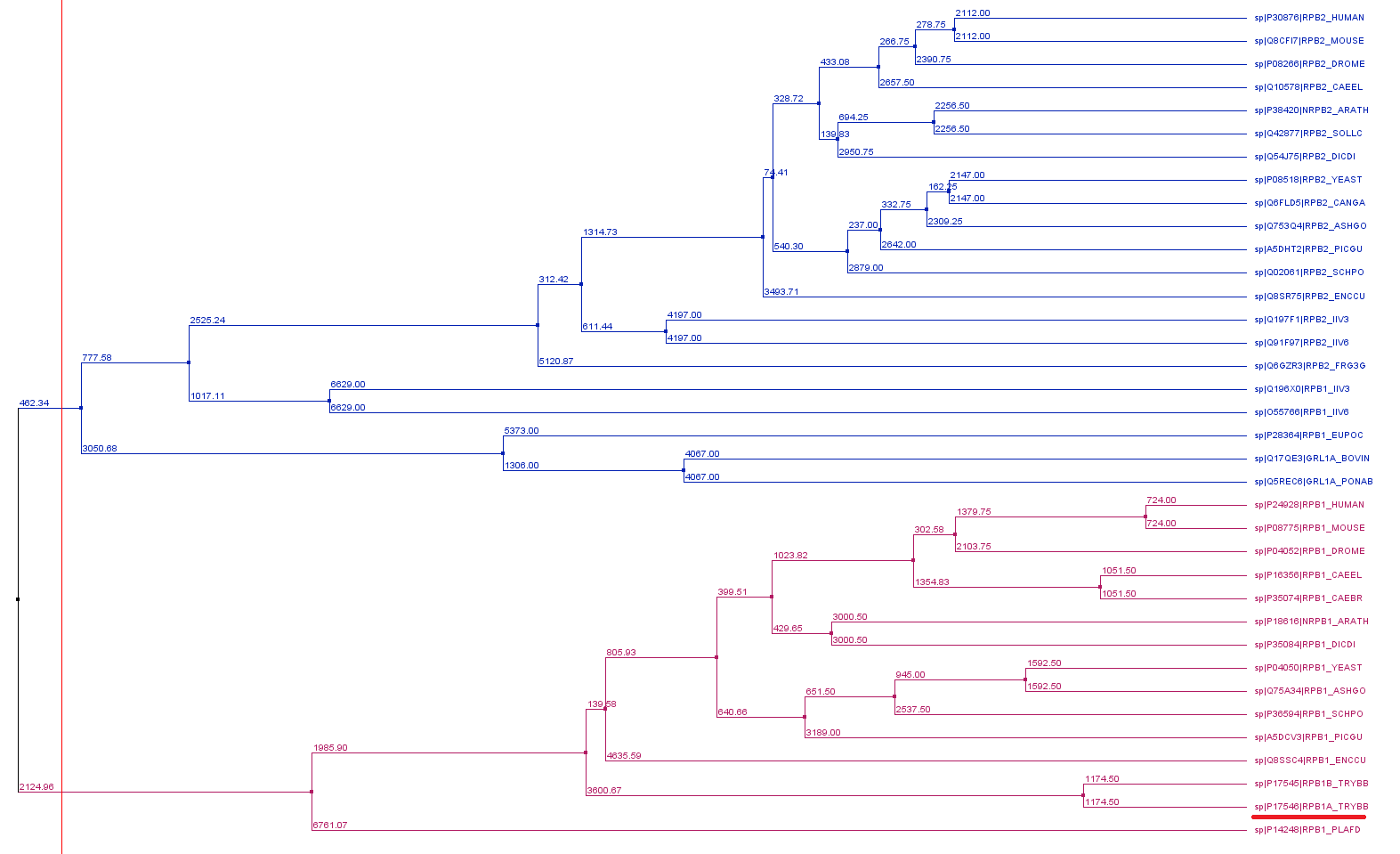
Phylogenetic reconstruction of Trypanosoma brucei brucei DdRPII RPB1 model DdRpII RPB1 protein sequences.
The tree was generated using the DdRpII family dataset (36 foul length protein sequences samples) and the Jalview software. Tree was constructed using the average distance statistical method with PAM 250. In the tree representation there are clearly shown the two RNA polymerases II subunits RPB1 and RPB2 as two main monophyletic sub-trees. Trypanosoma brucei DdRpII RPB1 protein sequence was correctly classified in the monophyletic sub-tree of the RPB1 group.
Multiple sequence alignment
The alignment was performed using the Trypanosoma brucei brucei DdRPII RPB1, the Trypanosoma brucei gambiense DdRpII RPB1, the crystal structure of Schizosaccharomyces pombe DdRpII RPB, the crystal structure of Saccharomyces cerevisiae DdRpII RPB1 and the electron microscopy structure of Human DdRpII DdRpII RPB1. (A) All nine suggested conserved motifs and major domains of DdRpII RPB1 have been marked (Motifs 1a, 1b, 2, 3a, 3b, 3c, 4a, 4b, 4c). Additionally, in the multiple sequence alignment were presented the major differences. (B) Domains and domainlike regions of the DdRpII subunit Rpb1. The amino acid residue numbers at the domain boundaries are indicated.
Multiple sequence alignment
The alignment was performed using the Trypanosoma brucei brucei DdRPII RPB1, the crystal structure of Schizosaccharomyces pombe DdRpII RPB and the electron microscopy structure of Bos taurus DdRpII RPB1. All five sub-domains (A-E) as referred in Pfam database have been marked with different colours.
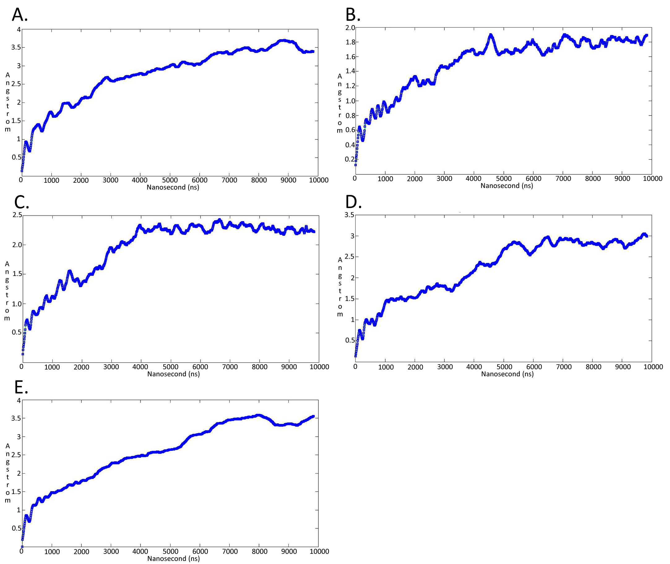
Molecular dynamicssimulation charts of the root mean square deviation (RMSD) for the Trypanosoma brucei brucei DdRpII RPB1 sub domainsof the model A.
The energy (Kcal/mol) vs time (ns) plot of the 100ns simulation trajectory of the TBB DdRpII RPBI model A. Sub-domain regions of the Trypanosoma brucei brucei DdRPII RPB1 have been separated according to conventions of Supplementary Figure 3. (A) Domain A RMSD. (B) Domain B RMSD. (C) Domain C RMSD. (D) Domain D RMSD. (E) Domain E RMSD.
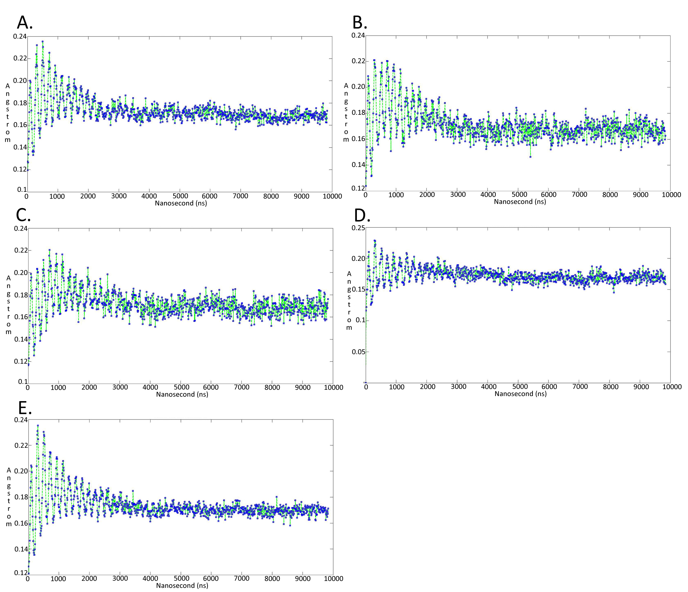
Molecular dynamics simulationcharts of the root mean square fluctuation (RMSF) for the Trypanosoma brucei brucei DdRpII RPB1 sub domains of the model A.
Sub-domain regions of the Trypanosoma brucei brucei DdRPII RPB1 have been separated according to conventions of Supplementary Figure 3. (A) Domain A RMSF. (B) Domain B RMSF. (C) Domain C RMSF. (D) Domain D RMSF. (E) Domain E RMSF.
Molecular dynamics simulationcharts of the root mean square deviation (RMSD) for the Trypanosoma brucei brucei DdRpII RPB1 sub domainsof the model B.
The energy (Kcal/mol) vs time (ns) plot of the 100ns simulation trajectory of the TBB DdRpII RPBI model B. Sub-domain regions of the Trypanosoma brucei brucei DdRPII RPB1 have been separated according to conventions of Supplementary Figure 3. (A) Domain A RMSD. (B) Domain B RMSD. (C) Domain C RMSD. (D) Domain D RMSD. (E) Domain E RMSD.
Molecular dynamics simulationcharts of the root mean square fluctuation (RMSF) for the Trypanosoma brucei brucei DdRpII RPB1 sub domains of the model B.
Sub-domain regions of the Trypanosoma brucei brucei DdRPII RPB1 have been separated according to conventions of Supplementary Figure 3. (A) Domain A RMSF. (B) Domain B RMSF. (C) Domain C RMSF. (D) Domain D RMSF. (E) Domain E RMSF.
MEGA software phylogenetic tree in newick format
The tree was constructed the Neighbour – Joining statistical method for 100 bootstrap replicates and the 36 extracted samples of the DpRpII.
Supplementary Data 3: Jalview software phylogenetic tree in newick format
The tree was constructed using the average distances statistical method and the 36 extracted samples of the DpRpII.
Trypanosoma brucei brucei DdRPII RPB1 model A in .pdb format.
Trypanosoma brucei brucei DdRPII RPB1 model B in .pdb format.