Chemical modulation of apoptosis in molluscan cell cultures
- Published
- Accepted
- Subject Areas
- Cell Biology, Marine Biology, Molecular Biology
- Keywords
- : Apoptotic inducers, Apoptotic inhibitors, Cell death pathways, Flow cytometry, Mussel, Mytilus trossulus
- Copyright
- © 2018 Boroda et al.
- Licence
- This is an open access article distributed under the terms of the Creative Commons Attribution License, which permits unrestricted use, distribution, reproduction and adaptation in any medium and for any purpose provided that it is properly attributed. For attribution, the original author(s), title, publication source (PeerJ Preprints) and either DOI or URL of the article must be cited.
- Cite this article
- 2018. Chemical modulation of apoptosis in molluscan cell cultures. PeerJ Preprints 6:e27100v1 https://doi.org/10.7287/peerj.preprints.27100v1
Abstract
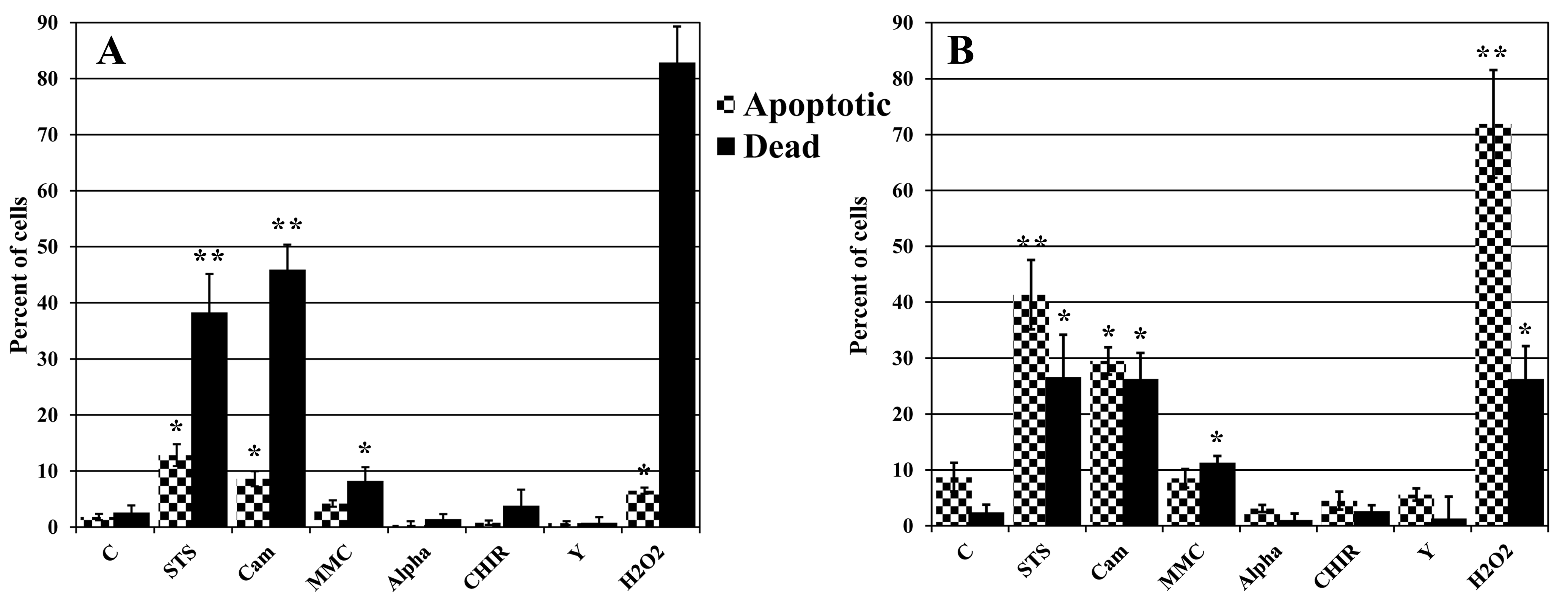
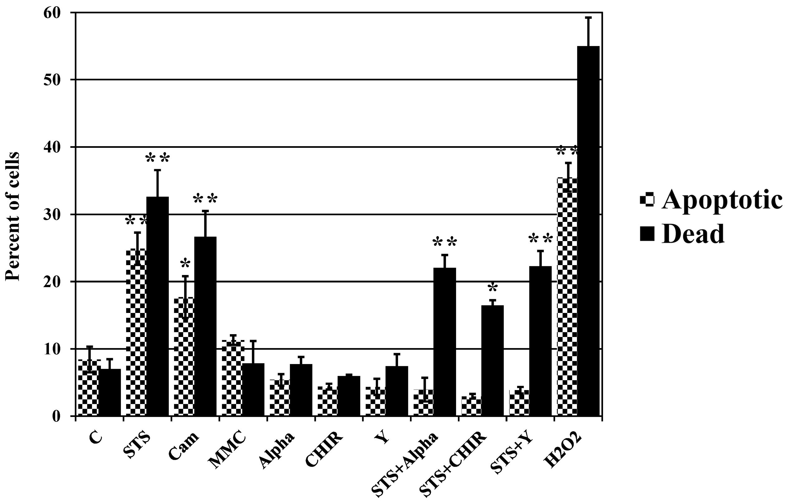
This study focused on the alterations that occur in larval molluscan cells during the induction or inhibition of apoptosis both in standard culture conditions and in response to cold injury during the induction of different death pathways. This is the first report on the modulation of apoptosis in molluscan cells using apoptotic inducers and inhibitors known to mammalian cells, which has been assessed by flow cytometry. The activity of mitochondria, general caspase activation, and the membrane integrity of intact molluscan cells were compared to those of cells frozen-thawed both prior to treatment and after incubation with apoptotic inducers or inhibitors, and to those of primary mouse embryonic fibroblasts and human colon tumor cells (HCT 116 cell line) treated with the same compounds. We tested three apoptotic inducers (staurosporine, camptothecin, and mitomycin C, routinely used for the chemical induction of apoptosis in different mammalian cells) and found that only staurosporine resulted in an evident increase of apoptosis in molluscan cells (6.6% in comparison with 2.9% in control unfrozen cells, and 9.1% in comparison with 5.6% in control frozen-thawed cells). Camptothecin did not significantly induce apoptosis of molluscan cells but did slightly increase the number of active cells after thawing. Mitomycin C showed similar results, but its effect was less pronounced. We suggest that some apoptotic inducers have hereto unknown effects on molluscan cells. In addition, we hypothesize that the use of the apoptotic inhibitors could reduce apoptosis, which is significant after cryopreservation in molluscan cells. Development of this direction is important for understanding the mechanisms of cold susceptibility of marine organisms.
Author Comment
This is a submission to PeerJ for review.