Mapping the self-association domains of ataxin-1: Identification of novel non overlapping motifs
- Published
- Accepted
- Subject Areas
- Biochemistry, Cell Biology
- Keywords
- polyglutamine diseases, confocal microscopy, spinocerebellar ataxia type, FRET, foci, misfolding diseases
- Copyright
- © 2014 Menon et al.
- Licence
- This is an open access article distributed under the terms of the Creative Commons Attribution License, which permits unrestricted use, distribution, reproduction and adaptation in any medium and for any purpose provided that it is properly attributed. For attribution, the original author(s), title, publication source (PeerJ PrePrints) and either DOI or URL of the article must be cited.
- Cite this article
- 2014. Mapping the self-association domains of ataxin-1: Identification of novel non overlapping motifs. PeerJ PrePrints 2:e259v2 https://doi.org/10.7287/peerj.preprints.259v2
Abstract
The neurodegenerative spinocerebellar ataxia type 1 (SCA1) is caused by aggregation and misfolding of the ataxin-1 protein. While the pathology correlates with mutations that lead to expansion of a polyglutamine tract in the protein, other regions contribute to the aggregation process as also non-expanded ataxin-1 is intrinsically aggregation-prone and forms nuclear foci in cell. Here, we have used a combined approach based on FRET analysis, confocal microscopy and in vitro techniques to map aggregation-prone regions other than polyglutamine and to establish the importance of dimerization in self-association/foci formation. Identification of aggregation-prone regions other than polyglutamine could greatly help the development of SCA1 treatment more specific than that based on targeting the low complexity polyglutamine region.
Supplemental Information
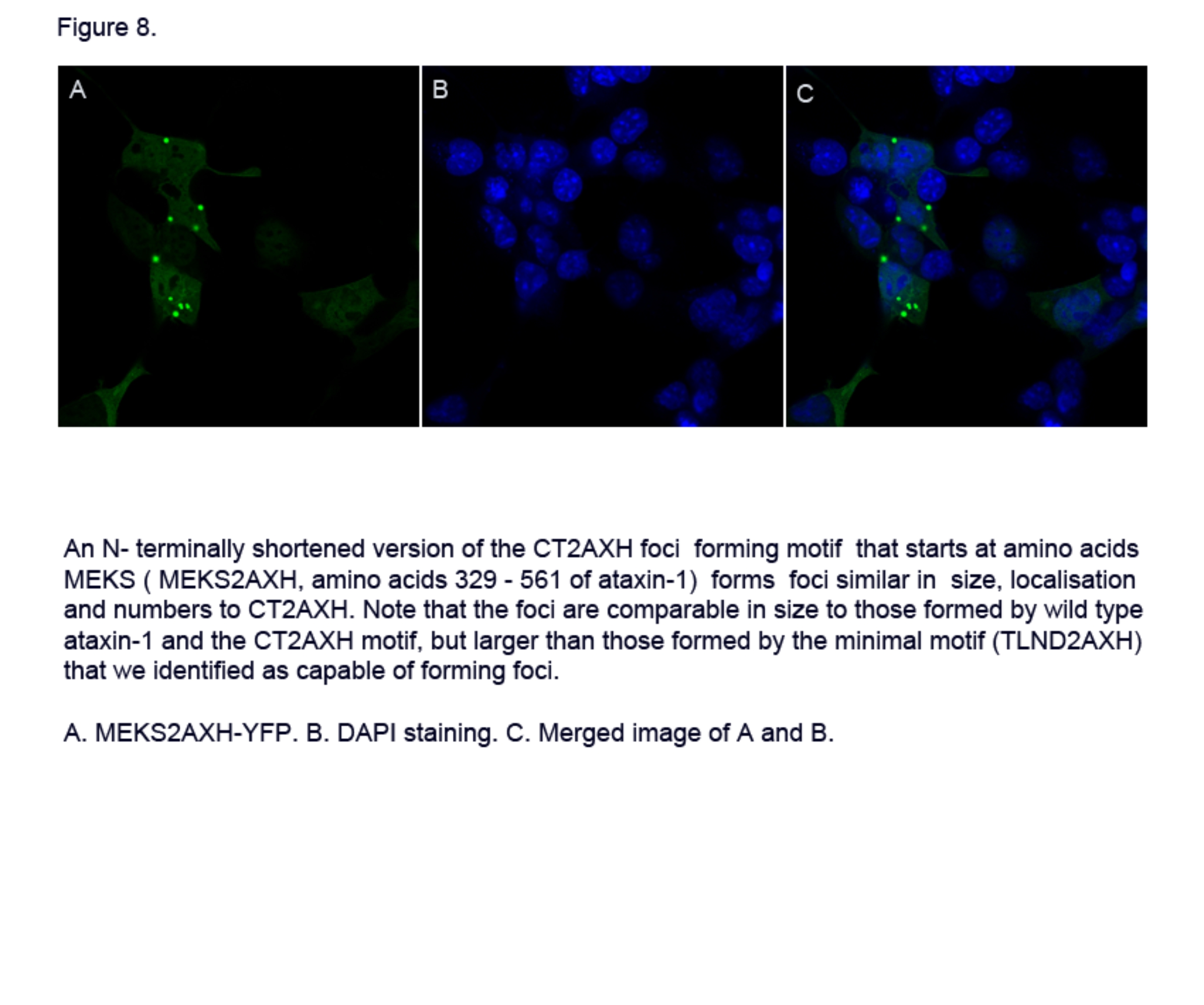
Figure 8 - Foci formation of the shorter construct MEKS2AXH, aa 329-561 of ataxin-1.
Figure 8 - Foci formation of the shorter construct MEKS2AXH, aa 329-561 of ataxin-1.