The reappraisal of Batson’s theory by isolated para-vertebral metastasis of hepatocellular carcinoma
- Published
- Accepted
- Subject Areas
- Radiology and Medical Imaging
- Keywords
- Hepatocellular carcinoma, metastasis
- Copyright
- © 2014 Liang et al.
- Licence
- This is an open access article distributed under the terms of the Creative Commons Attribution License, which permits unrestricted use, distribution, and reproduction in any medium, provided the original author and source are credited.
- Cite this article
- 2014. The reappraisal of Batson’s theory by isolated para-vertebral metastasis of hepatocellular carcinoma. PeerJ PrePrints 2:e212v1 https://doi.org/10.7287/peerj.preprints.212v1
Abstract
The extra-hepatic metastasis of hepatocellular carcinoma (HCC) usually hints late disease stage and poor prognosis. However, we treated 5 unusual HCC patients with isolated para-vertebral metastasis by tumor excision and the surgical result is satisfactory during 3.5 years follow-up in mean. To explain why the unusual situation of isolated metastasis of HCC occurred, an alternative spreading route of cancer cells bypass pulmonary circulation and avoid pulmonary vascular filtration, the Batson’s plexus, was highly suspected. We supposed that isolated para-vetebral metastases seems not to mean a late disease stage, aggressive management made longer survival possible, and it was confirmed by our clinical experience.
Supplemental Information
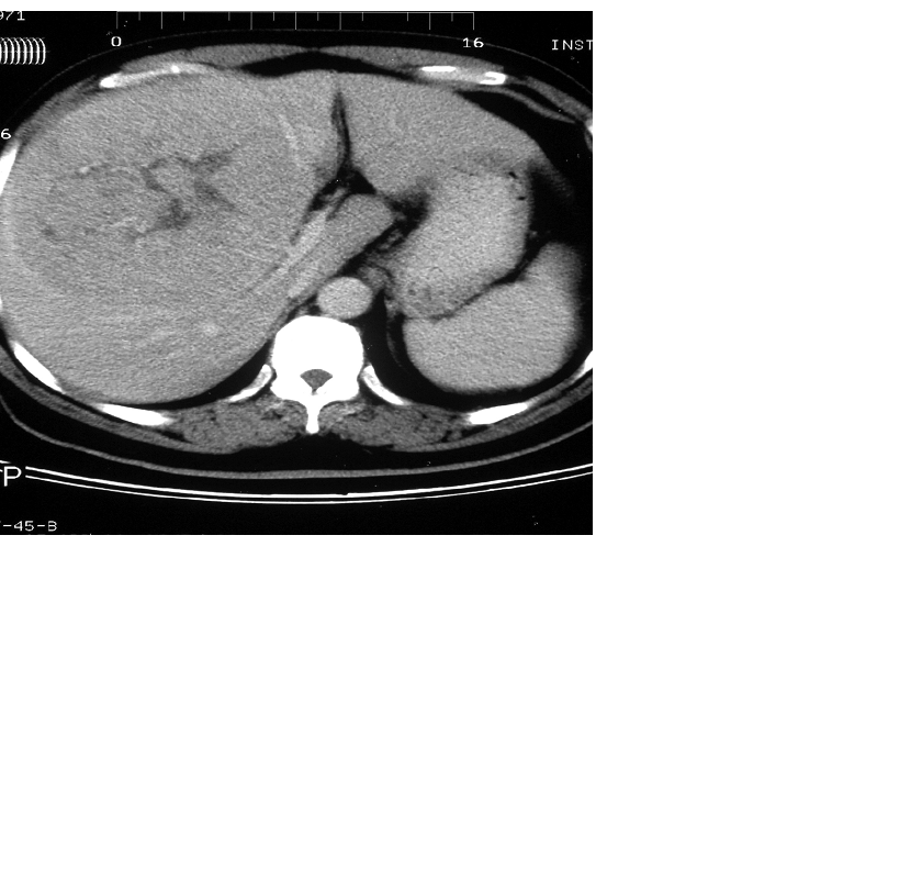
The abdominal CT scan for primary liver tumor.
A 10.6 x 8.8 cm hepatic tumor was noted over right lobe by contrast-enhanced CT.
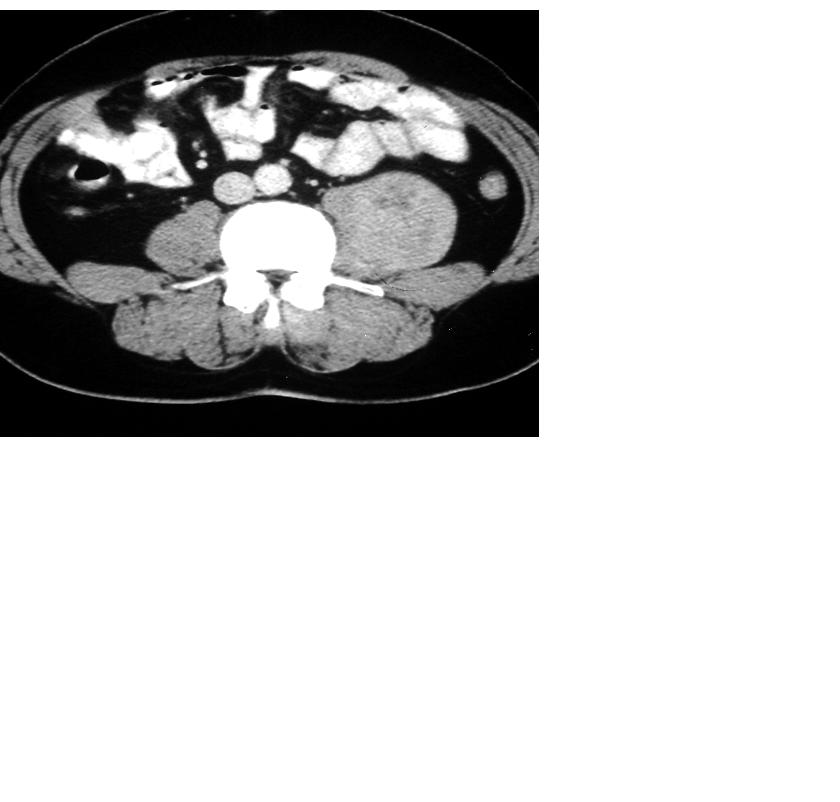
The abdominal CT scan for metastatic lesion.
The heterogeneous mass over left psoas muscle was noted by contrast-enhanced CT, the size is 4x4x 3 cm (white arrow).
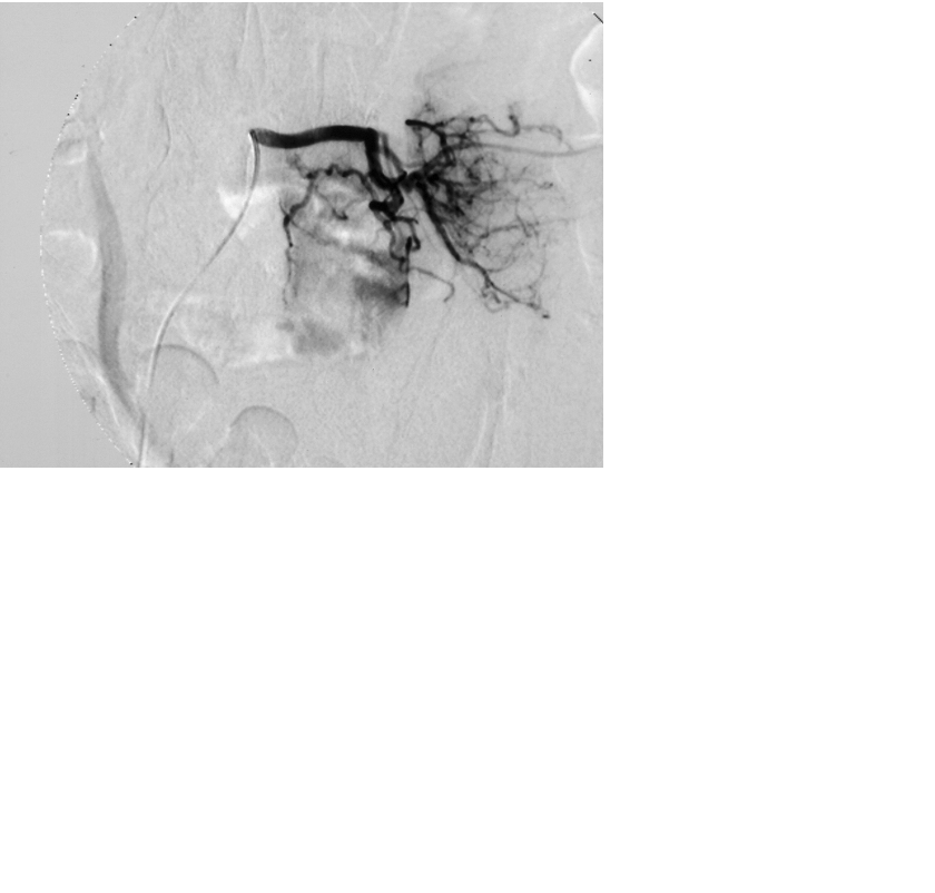
The abdominal CT scan for metastatic lesion.
Hypervascularity of the psoas tumor was proved by abdominal angiography(white arrow).
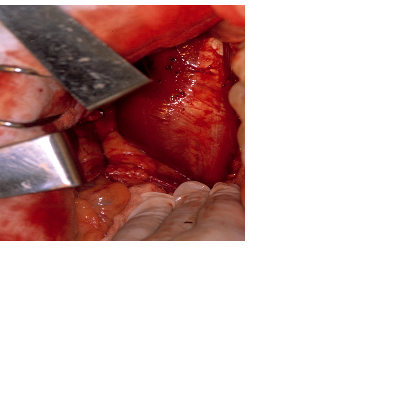
The gross picture of metastatic lesion.
One fragile, reddish mass within the psoas muscle was observed during operation(white arrow).
The abdominal CT scan for metastatic lesion
The right para-vertebral mass showed the characteristics of high signal in MRI image (white arrow).
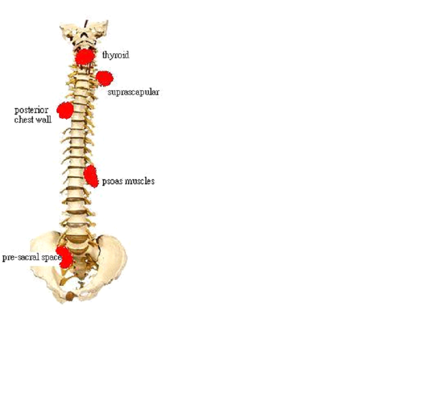
The metastatic sites noted over para-vertebral area.
The metastasis locations (red spot) hint the presence of metastatic route via Batson’s venous.