Preliminary evidence of a new microbial species capable of sustainable intracellular survival and transfer in mammalian cell lines
- Published
- Accepted
- Subject Areas
- Biochemistry, Cell Biology, Molecular Biology
- Keywords
- quality, microbe, sterility, intracellular contamination, Tissue culturecontamination, Black dot swiming in tissue culture, Black dot
- Copyright
- © 2014 Barkwan et al.
- Licence
- This is an open access article distributed under the terms of the Creative Commons Attribution License, which permits unrestricted use, distribution, and reproduction in any medium, provided the original author and source are credited.
- Cite this article
- 2014. Preliminary evidence of a new microbial species capable of sustainable intracellular survival and transfer in mammalian cell lines. PeerJ PrePrints 2:e209v2 https://doi.org/10.7287/peerj.preprints.209v2
Abstract
The minimisation of exposure of mammalian cell lines to potential microbial contaminants is handled by routine adherence to quality laboratory procedures. Mycoplasma, are capable of sustainable intracellular existence, are not v isible in light microscopy and must be tested for using dedicated methods. Bacterial contamination is usually detectable by relatively simple optical, spectroscopy and pH methodology. Symbiont occupation assumes an evolved mutually beneficial relationship and does occur with many eukaryote-prokaryotes, but rarely mammals. Other, purely intracellular low density, low energy and relatively stable and non-visible bacterial occupation of mammalian cytoplasm, assumes the existence of new intra-genus relationships and associated mechanisms. In this study, preliminary microscopy and sequence data has been collated implying the presence of low density Cocci in the cytoplasm of hepatocyte lines with negligible impact on cell function and behaviour.
Author Comment
I did few simple changes listed below: 1. Remove my name, email address and phone number under title sentence. 2. Remove the line numbers. 3. Change the color for some words to black as rest of the text. e.g. Fig. 4. Editing the (font face) of the Reference.
Supplemental Information
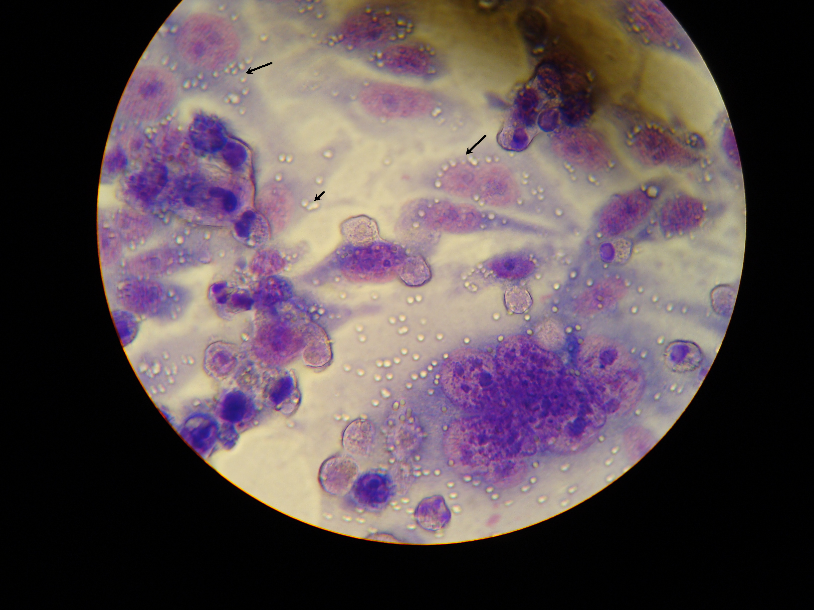
Giemsa stained cells
The arrow in the Light microscope image showing ‘transparent’ round shape bodies observed adjacent to lysed Giemsa stained cells while the nucleus and cytoplasm are stained with purple and pink colour (x200). Image restricted by condenser to maximise core illumination.
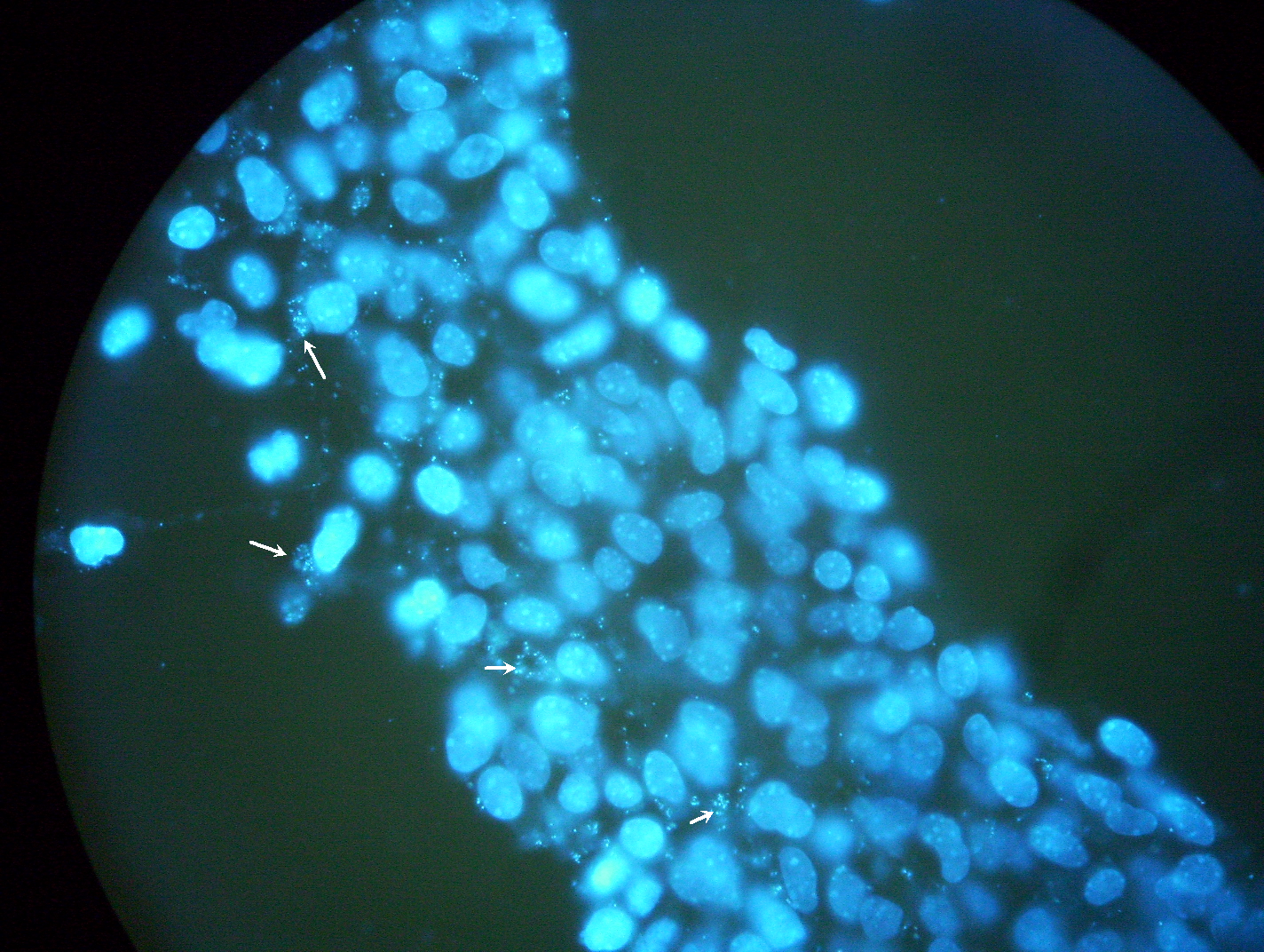
DAPI Stain
Fixed cells on cover slips and stained with counterstain DAPI. Arrow point to the Spots of nuclear materials observed around the nucleus of cells in the image (x100).
TEM micrographs of C3A cells infected with unknown bacteria
Bacteria are engulfed by elongated microvilli from infected epithelial cells. High magnification showing partially lysed vacuole membrane containing bacteria, indicating the ability of bacteria to escape from the endocytic vacuoles.
TEM micrographs of C3A cells infected with unknown bacteria (~ 1 m m dia).
(~ 1 m m dia). Cross section of the cells monolayer showing numerous intracellular bacteria. Micrograph showing membrane ‘ruffling’ upon contact with bacteria.