Altered ADAR1 in mice affected by social isolation stress-induced cognitive deficits
- Published
- Accepted
- Subject Areas
- Animal Behavior, Biochemistry, Neuroscience
- Keywords
- cognitive ability, ADAR1, social isolation
- Copyright
- © 2016 Chen et al.
- Licence
- This is an open access article distributed under the terms of the Creative Commons Attribution License, which permits unrestricted use, distribution, reproduction and adaptation in any medium and for any purpose provided that it is properly attributed. For attribution, the original author(s), title, publication source (PeerJ Preprints) and either DOI or URL of the article must be cited.
- Cite this article
- 2016. Altered ADAR1 in mice affected by social isolation stress-induced cognitive deficits. PeerJ Preprints 4:e1870v1 https://doi.org/10.7287/peerj.preprints.1870v1
Abstract
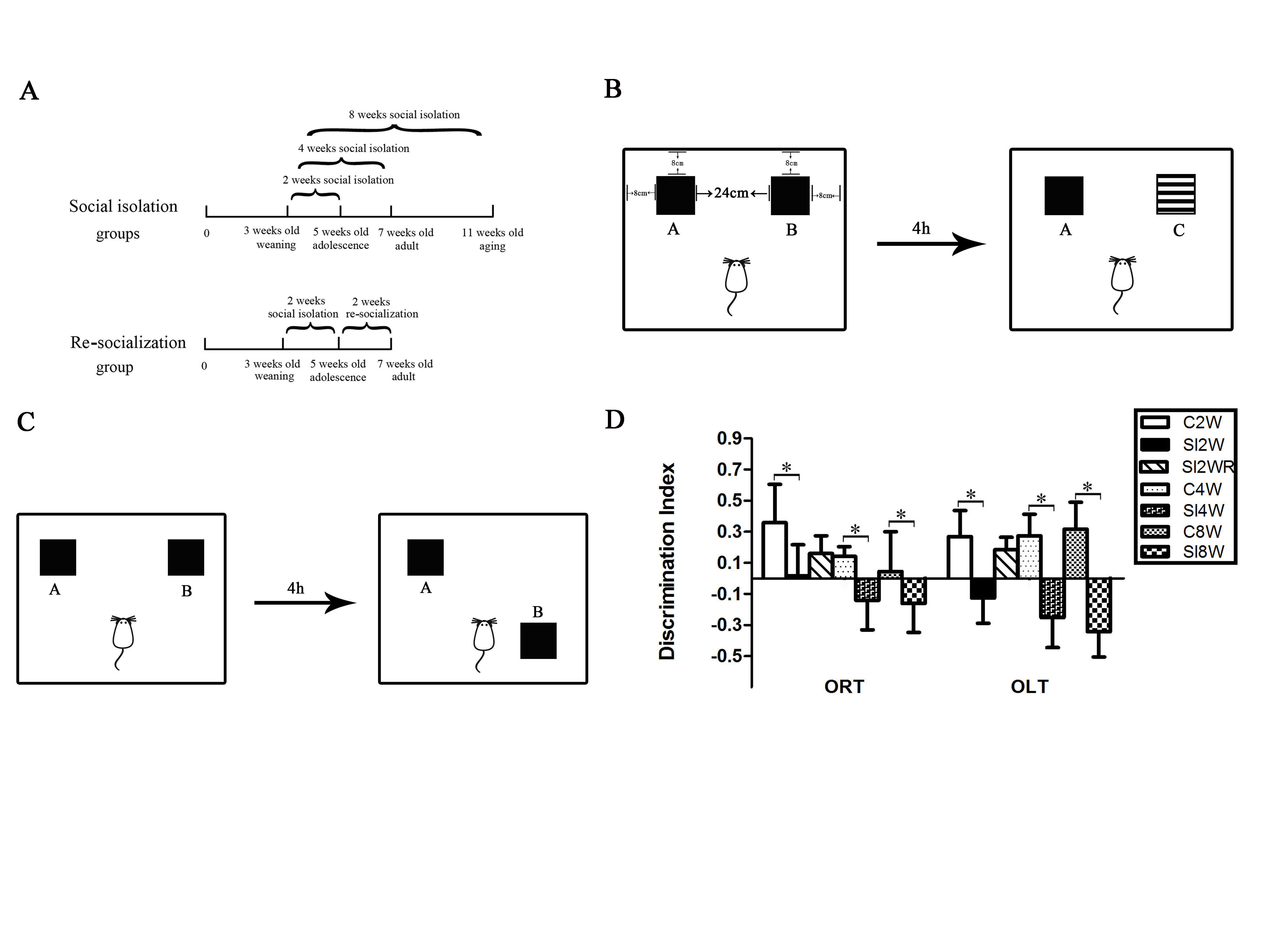
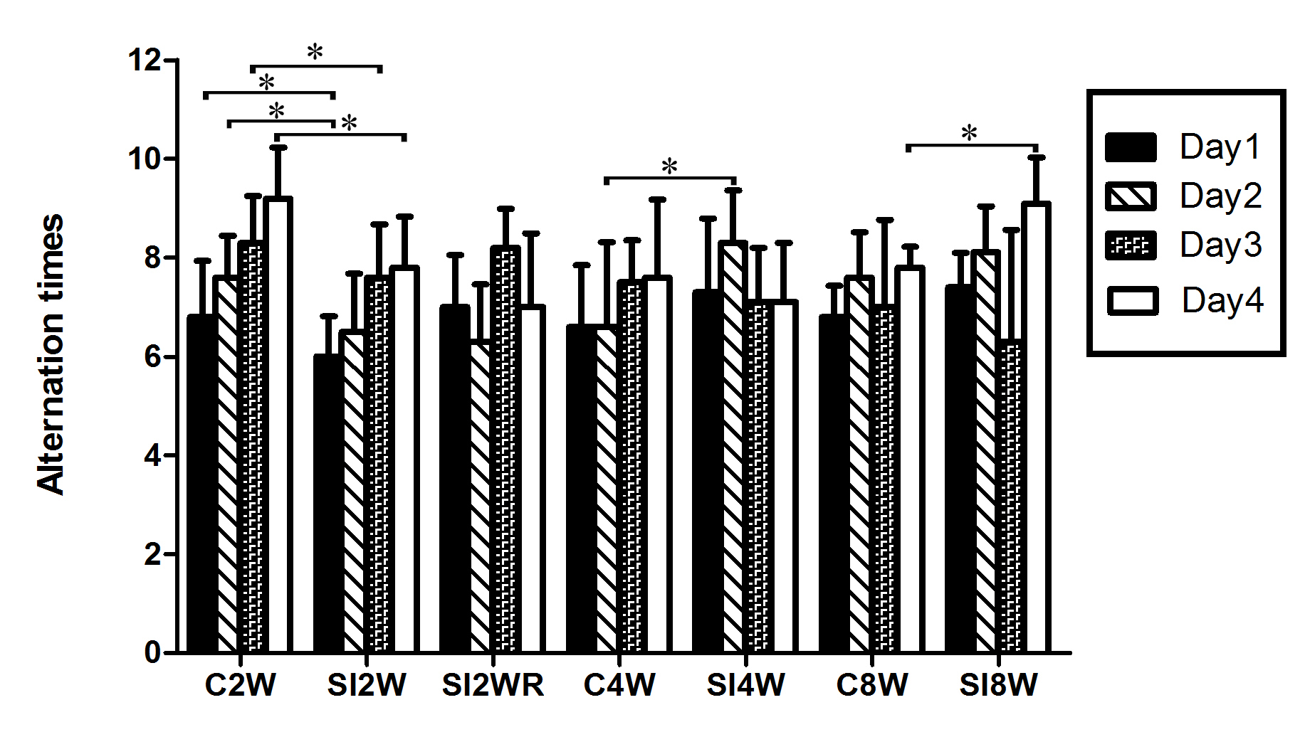
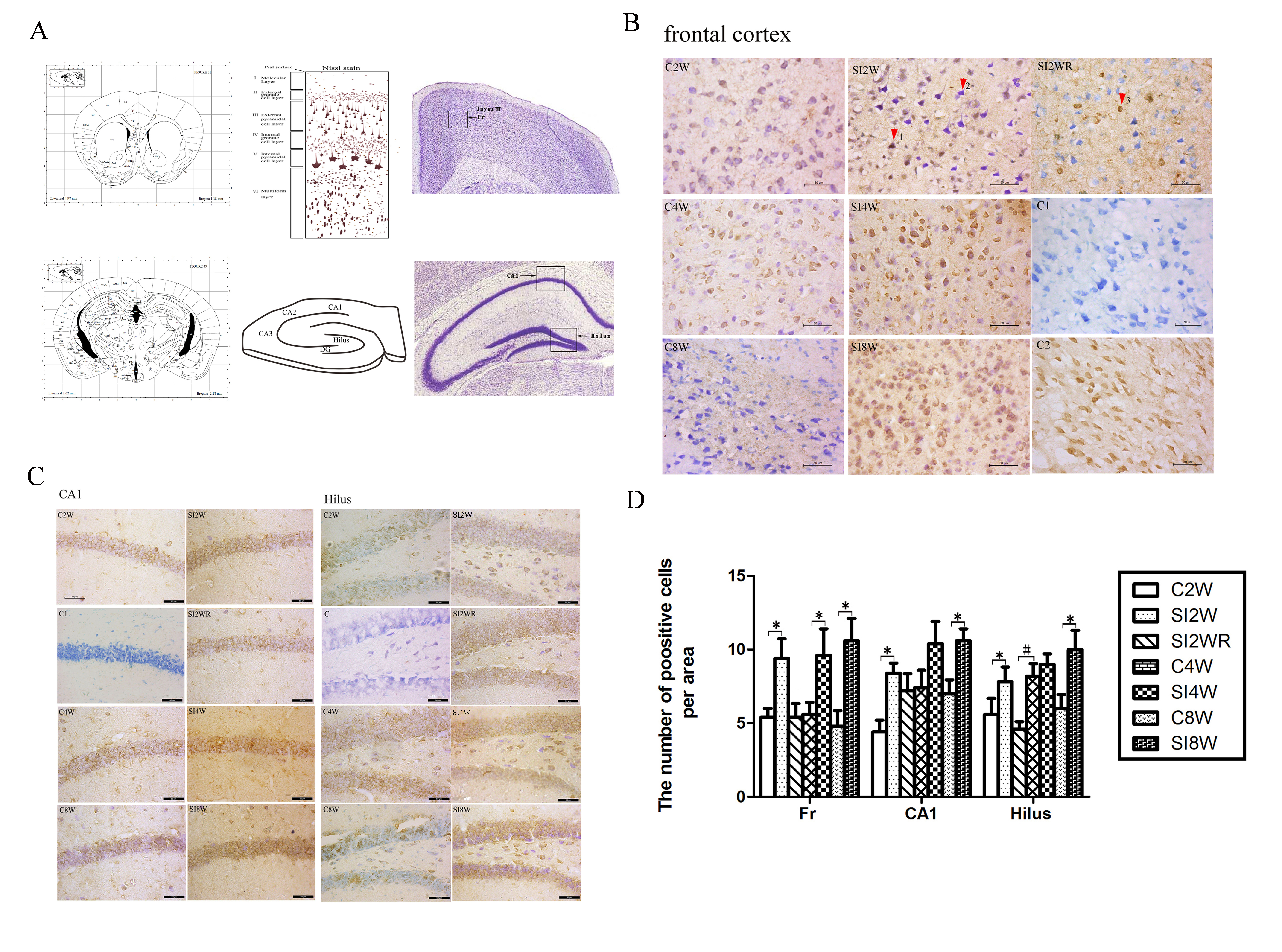
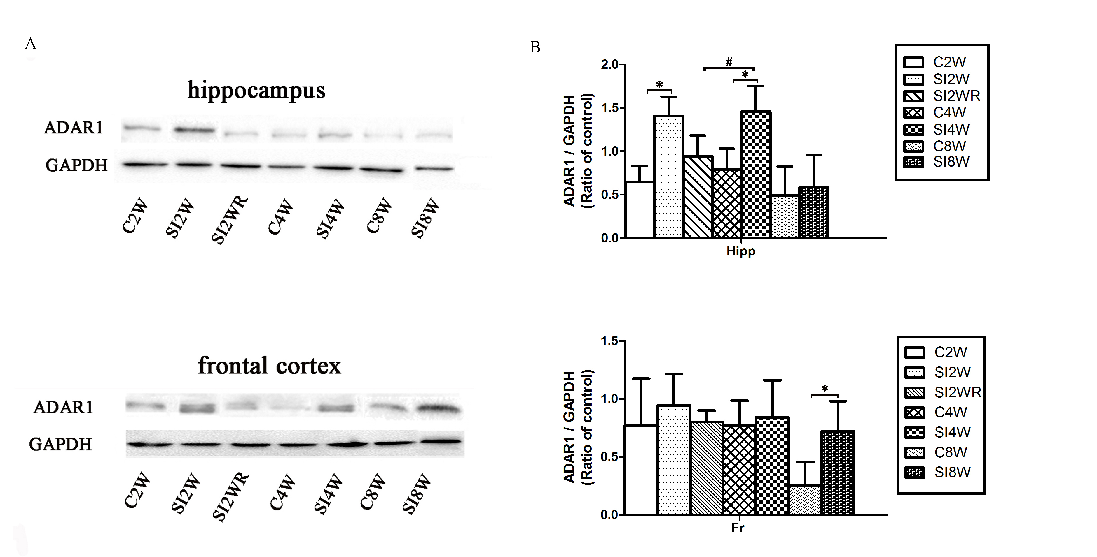
Adenosine deaminase acting on RNA (ADAR) activity increases in response to inflammation. Social isolation stress is related to neuroinflammation; however, it remains unclear whether ADAR1 is altered in response to social isolation stress-induced cognitive deficits. To investigate our hypothesis that ADAR1 displayed patterns of change in response to social isolation stress, we addressed this issue systemically by isolating Kunming mice for 2, 4 and 8 weeks individually since postnatal 21 days to set up isolation mouse model. Furthermore, we arranged re-socialization group to evaluate the alterations of ADAR1 in the cognitive deficits recovery. The results of behavior tests showed that social isolation stress resulted in cognitive dysfunction, which was recovered by re-socialization in re-gregarious rearing group. Furthermore, the immunohistochemistry and western blot results displayed that both the immunoreactivity and protein expression of ADAR1 in the hippocampus and frontal cortex increased obviously as compared to the same age mice without isolation. The above abnormal alterations of ADAR1 were recovered by re-socialization in re-gregarious rearing group. Our study supports the hypothesis that ADAR1 is altered in mice affected by social isolation stress-induced cognitive deficits.
Author Comment
This is a preprint submission to PeerJ PrePrints.