A preliminary case study of the effect of shoe-wearing on the biomechanics of a horse’s foot
- Published
- Accepted
- Subject Areas
- Animal Behavior, Veterinary Medicine, Zoology
- Keywords
- equine biomechanics, farriery, finite element analysis, inverse dynamics, XROMM, locomotion, horse foot
- Copyright
- © 2016 Panagiotopoulou et al.
- Licence
- This is an open access article distributed under the terms of the Creative Commons Attribution License, which permits unrestricted use, distribution, reproduction and adaptation in any medium and for any purpose provided that it is properly attributed. For attribution, the original author(s), title, publication source (PeerJ PrePrints) and either DOI or URL of the article must be cited.
- Cite this article
- 2016. A preliminary case study of the effect of shoe-wearing on the biomechanics of a horse’s foot. PeerJ PrePrints 4:e1779v1 https://doi.org/10.7287/peerj.preprints.1779v1
Abstract
Horse racing is a multi-billion-dollar industry that has raised welfare concerns due to disabled and euthanized animals. Whilst the cause of musculoskeletal injuries that lead to horse morbidity and mortality is multifactorial, pre-existing pathologies, increased speeds and substrate of the racecourse are likely contributors to foot disease. The hooves of horses have the ability to naturally deform during locomotion and dissipate locomotor stresses, yet farriery approaches are utilised to increase performance and protect hooves from wear. Previous studies have assessed the effect of different shoe designs on locomotor performance; however, no biomechanical study has hitherto measured the effect of horseshoes on the stresses of the foot skeleton in vivo. As there is a need to reduce musculoskeletal injuries in racing and training horses, it is crucial to understand the natural function of the feet of horses and how this is influenced by shoe design. This preliminary study introduces a novel combination of three-dimensional data from biplanar radiography, inverse dynamics, and finite element analysis (FEA) to evaluate the effect of a stainless steel shoe on the function of a Thoroughbred horse’s front foot during walking. Our results show that the stainless steel shoe increases craniocaudal, mediolateral and vertical GRFs at mid-stance. We document a similar pattern of flexion-extension in the PIP (pastern) and DIP (coffin) joint between the unshod and shod conditions, yet variation in the degrees of rotations are encountered throughout the stance phase. In particular, in both the shod and unshod conditions, the PIP joint extends between the 10-40% of the stance phase and flexes before mid-stance and until the end of the stance phase. Similarly the DIP joint extends until the 40% of stance and then flexes until the end of the stance phase. Overall at mid-stance the PIP joint extends more at the shod (-2.9o) than the unshod (-1.5o) horse, whilst the DIP joint extends more at the unshod (-3.6o), than the shod (-2.8o) condition. We also document that the DIP joint flexes more than the PIP after mid-stance and until the end of the stance in both conditions. Our FEA results show increased von Mises stresses on the fore foot phalanges in the shod condition at mid-stance, indicating that the steel shoe increases mechanical loading. Our preliminary study illustrates how the shoe may influence the dynamics and mechanics of a Thoroughbred horse’s forefoot during slow walking, but more research is needed to quantify the effect of the shoe on the equine forefoot during the whole stance phase, at faster speeds/gaits and with more individuals as well as with a similar focus on the hind feet. We anticipate that our preliminary analysis using advanced methodological approaches will pave the way for new directions in research on the form/function relationship of the equine foot, with the ultimate goal to minimise foot injuries and improve animal health and welfare.
Author Comment
This is a submission to PeerJ for review.
Supplemental Information
Raw speed data for the unshod (n=4) and shod (n=4) conditions
Column A shows the conditions. Column B lists the name and date of the steps. Column C lists the horse’s hip height in meters. Column D lists the frame rate of the Sony camera used for the speed calculations. Columns E and F list the start and end frame of each trial and each condition. Column G shows the difference between the start and end frame (i.e. number of frames elapsed). Column H shows the time in seconds and was calculated by dividing 1 over the camera frame rate (column D), multiplied by the frame difference (column G). Column I shows the distance that a marker placed on the middle of the body of the horse travelled between the start and end frames of the steps (columns E and F). Column J lists the velocity calculations per trial and condition. Velocity was measured by dividing the distance (column I) over the time (column H). Column K lists gravity at 9.81ms-2 and column L lists the Froude number per trial and condition. Rows J6 and J 12 show the average velocity for the unshod and shod condition respectively. Rows L6 and L12 show the average Froude number for the unshod and shod conditions respectively.
Ground reaction force (GRF) data in Newtons for the unshod (n=4) and shod (n=4) conditions
Degrees of motion for proximal interphalangeal (PIP) and distal interphalangeal (DIP) joints for the shod (n=4) and unshod (n=4) conditions about the flexion-extension axis
Raw von Mises stress data (MPa) for each condition and each bone segment
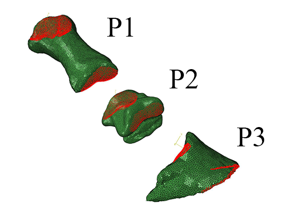
Bone segments are defined as the proximal phalanx (P1), intermediate phalanx (P2) and distal phalanx (P3). All stress data were exported from the external and internal nodes of the midshaft from homologous locations between bones and conditions as per Figure S3.
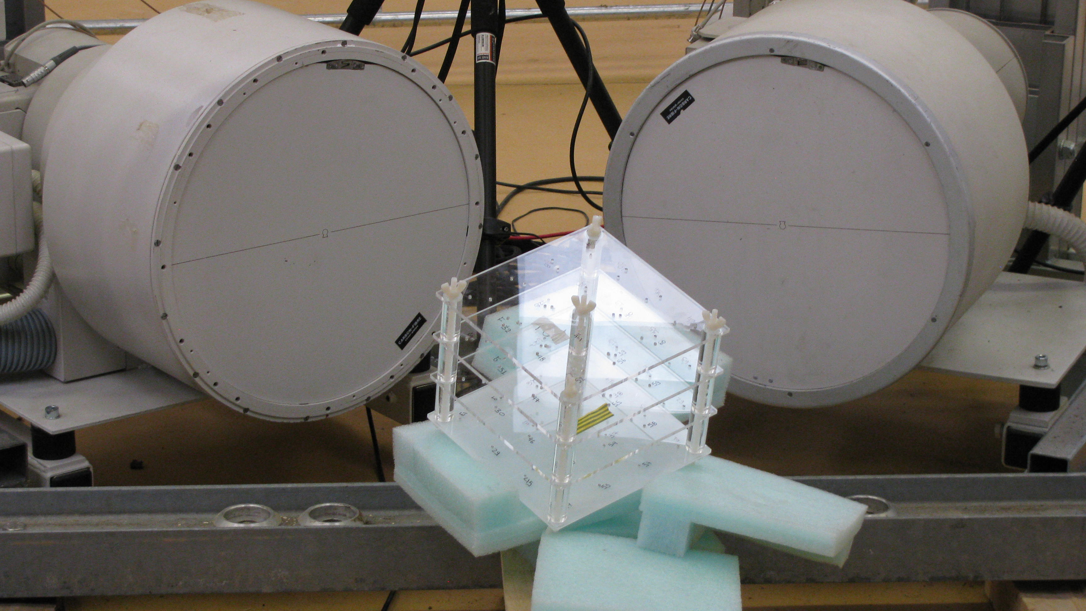
The position of the custom-designed calibration cube used during the fluoroscopy experiments to calibrate the 3D space in the XROMM analysis
Loading and boundary locations for the P1, P2 and P3 bones (see Methods: Loads and constraints)
Regional definitions (in red) for the P1 (A), P2 (B) and P3 (C) bones
All external and internal nodes of the midshaft were selected and nodal von Mises stresses were exported for the comparisons within homologous bones and both the shod and unshod conditions.