Cell cycle progression in glioblastoma cells is unaffected by pathophysiological levels of hypoxia
- Published
- Accepted
- Subject Areas
- Biochemistry, Cell Biology
- Keywords
- Hypoxia, Cell Cycle, HIF, Glioblastoma, Cell Death, Tumour Microenvironment, Brain Tumours
- Copyright
- © 2016 Richards et al.
- Licence
- This is an open access article distributed under the terms of the Creative Commons Attribution License, which permits unrestricted use, distribution, reproduction and adaptation in any medium and for any purpose provided that it is properly attributed. For attribution, the original author(s), title, publication source (PeerJ PrePrints) and either DOI or URL of the article must be cited.
- Cite this article
- 2016. Cell cycle progression in glioblastoma cells is unaffected by pathophysiological levels of hypoxia. PeerJ PrePrints 4:e1652v1 https://doi.org/10.7287/peerj.preprints.1652v1
Abstract
Hypoxia is associated with the increased malignancy of a broad range of solid tumours; however, whilst very severe hypoxia has been widely shown to induce cell cycle arrest, the impact of pathophysiological hypoxia on tumour cell proliferation is poorly understood. The aim of this study was to investigate the effect of different oxygen levels on glioblastoma (GBM) cell proliferation and survival. GBM is an extremely aggressive brain tumour with a heterogeneous oxygenation pattern. The effect of a range of oxygen tensions on GBM cell lines and primary cells were assessed using flow cytometry. Results indicate that cell cycle distribution and viability are unaffected by long term exposure (up to 4 days) to pathophysiological levels of oxygen (1-8% O 2). Both transient cell cycle arrest and small amounts of cell death could only be detected when cells were exposed to severe hypoxia (0.1% O2). No significant changes in p21 protein expression levels were detected. These findings reinforce the importance of using physiologically relevant oxygen tensions when investigating tumour hypoxia, and help to explain how solid tumours can be both hypoxic and highly proliferative, as is the case with GBM.
Author Comment
This is a submission to PeerJ for review.
Supplemental Information
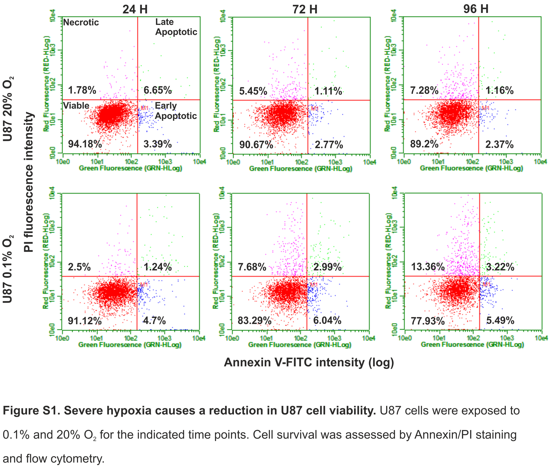
Severe hypoxia causes a reduction in U87 cell viability
U87 cells were exposed to 0.1% and 20% O2 for the indicated time points. Cell survival was assessed by Annexin/PI staining and flow cytometry.
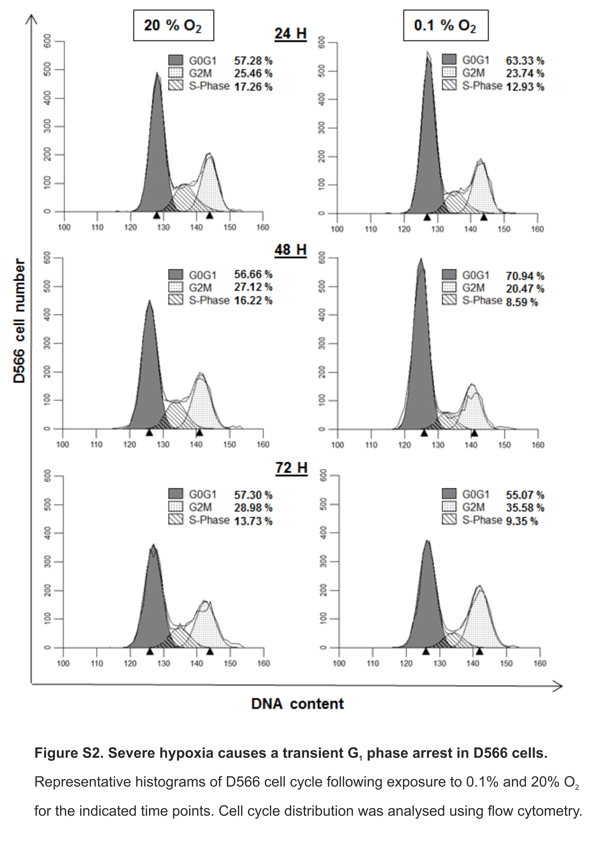
Severe hypoxia causes a transient G1 phase arrest in D566 cells
Representative histograms of D566 cell cycle following exposure to 0.1% and 20% O2 for the indicated time points. Cell cycle distribution was analysed using flow cytometry.
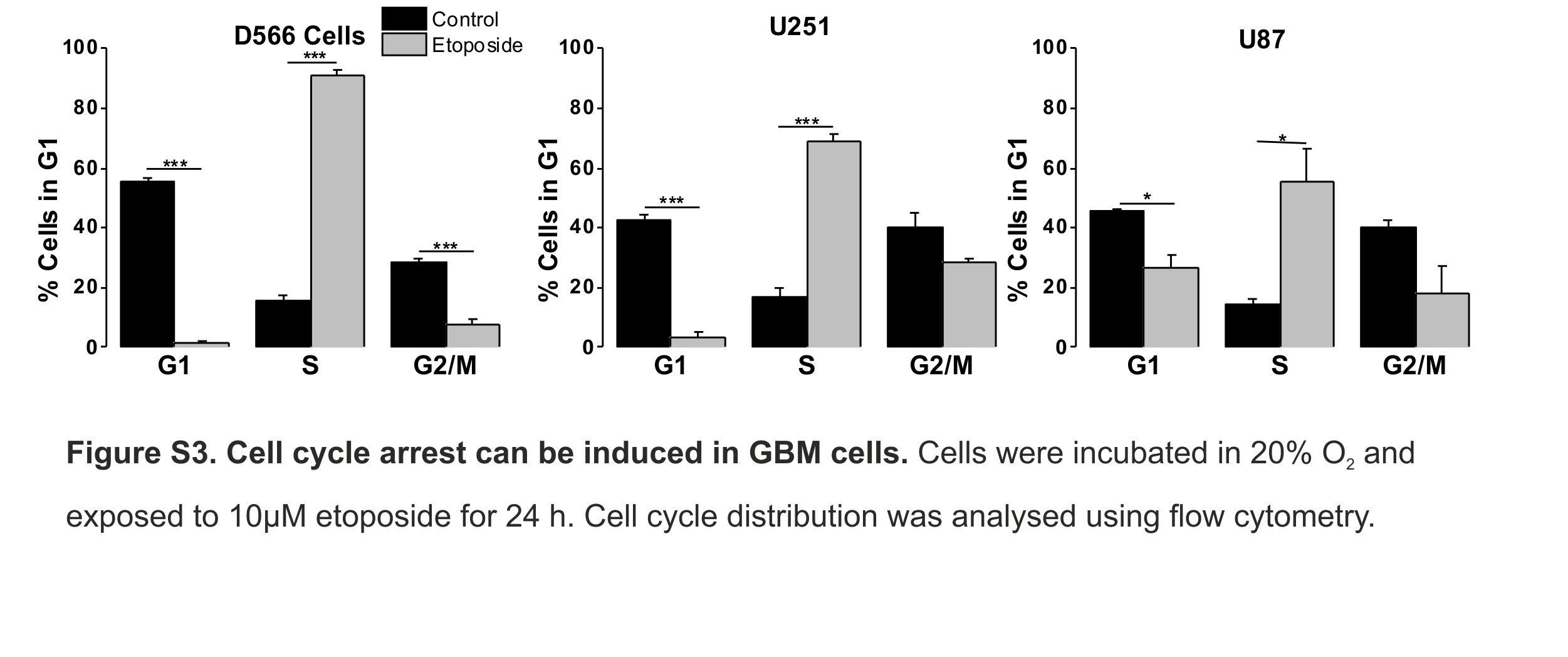
Cell cycle arrest can be induced in GBM cells
Cells were incubated in 20% O2 and exposed to 10µM etoposide for 24 h. Cell cycle distribution was analysed using flow cytometry.