
Following on from our past collaborations with the VIZBI conference series in 2019 and 2021, we sponsored two PeerJ Awards at the 12th International Meeting on Visualizing Biological Data, which took place virtually in March 2022.
VIZBI conferences bring together a diverse community, including bioinformaticians, data scientists, computer scientists, and experimentalists, as well as medical illustrators, graphic designers, and graphic artists. VIZBI 2022 featured talks from 21 world-leading researchers showcasing visualizations transforming how life scientists view data, and driving key advances in molecular biology, systems biology, biomedical science, and ecology.
We recently talked to the winners of PeerJ Awards for Best Scientific Poster and the Art & Biology Prize, with each also receiving a free publication (subject to peer review) in a PeerJ journal. If you are organising a conference and would like to discuss having a PeerJ Award at your meeting, please contact [email protected].
*****
Mol Mir Science Visualization Specialist, Stowers Institute for Medical Research.

Can you tell us a bit about yourself and your research interests?
I have two main research interests. The first is in understanding what cells look like in vivo. I work with planarians (flatworms that have mastered the art of regeneration thanks to their stem cells, called neoblasts) and serial block-face SEM. With this technology, we are able to view these stem cells as well as differentiating & differentiated cells in 3D at a very high resolution. My second research interest is in improving the ways we show & share data.
What first interested you in this field of research?
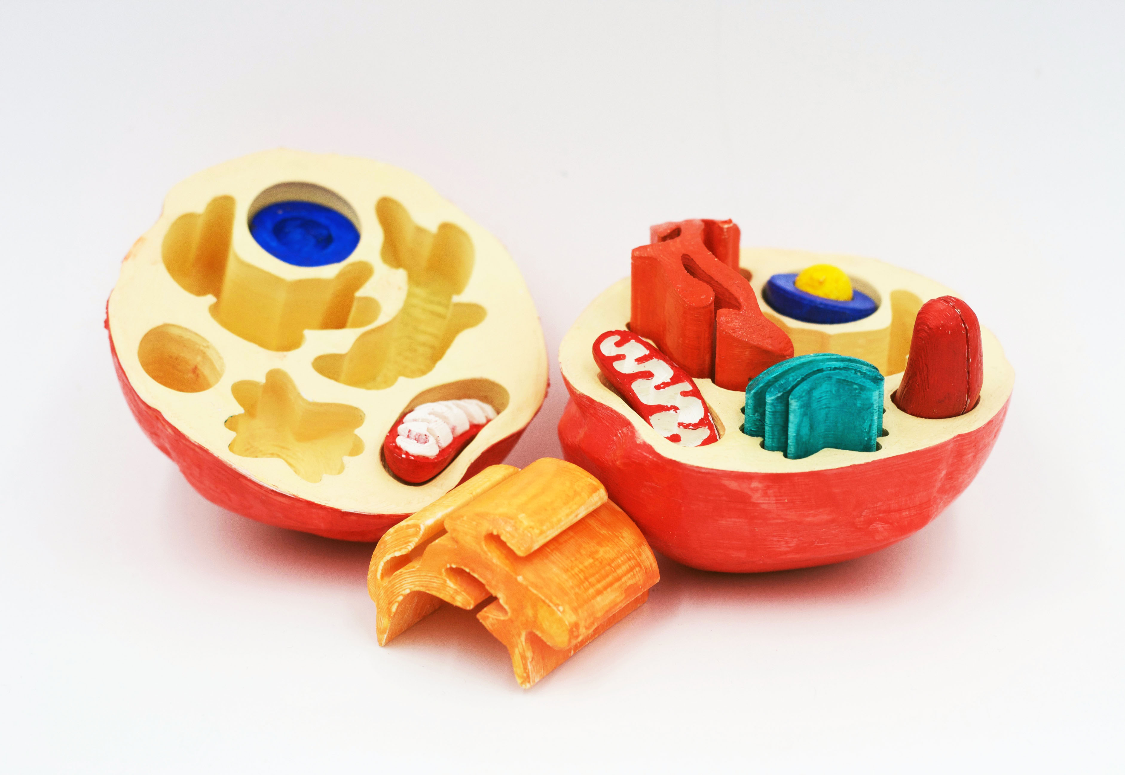
When I was in college, in the Interactive Arts program at Kansas City Art Institute, my first big 3D modeling project was an interactive 3D printed cell model for an intro biology class. I digitally sculpted the cell and organelles, including mitochondria that pull apart to reveal the cristae! This is where I began to have focused questions about what cells really look like. I got the opportunity to answer this question after a series of very fortunate events starting with a tour of Stowers Institute, resulting in an internship in the Sánchez Alvarado Lab. I immediately understood that there’s a dramatic disconnect between textbook illustrations of cells and microscopy images. I was – and still am – baffled that we continue to show such inaccurate and oversimplified representations of cells when we actually have incredible reference images of them in vivo.
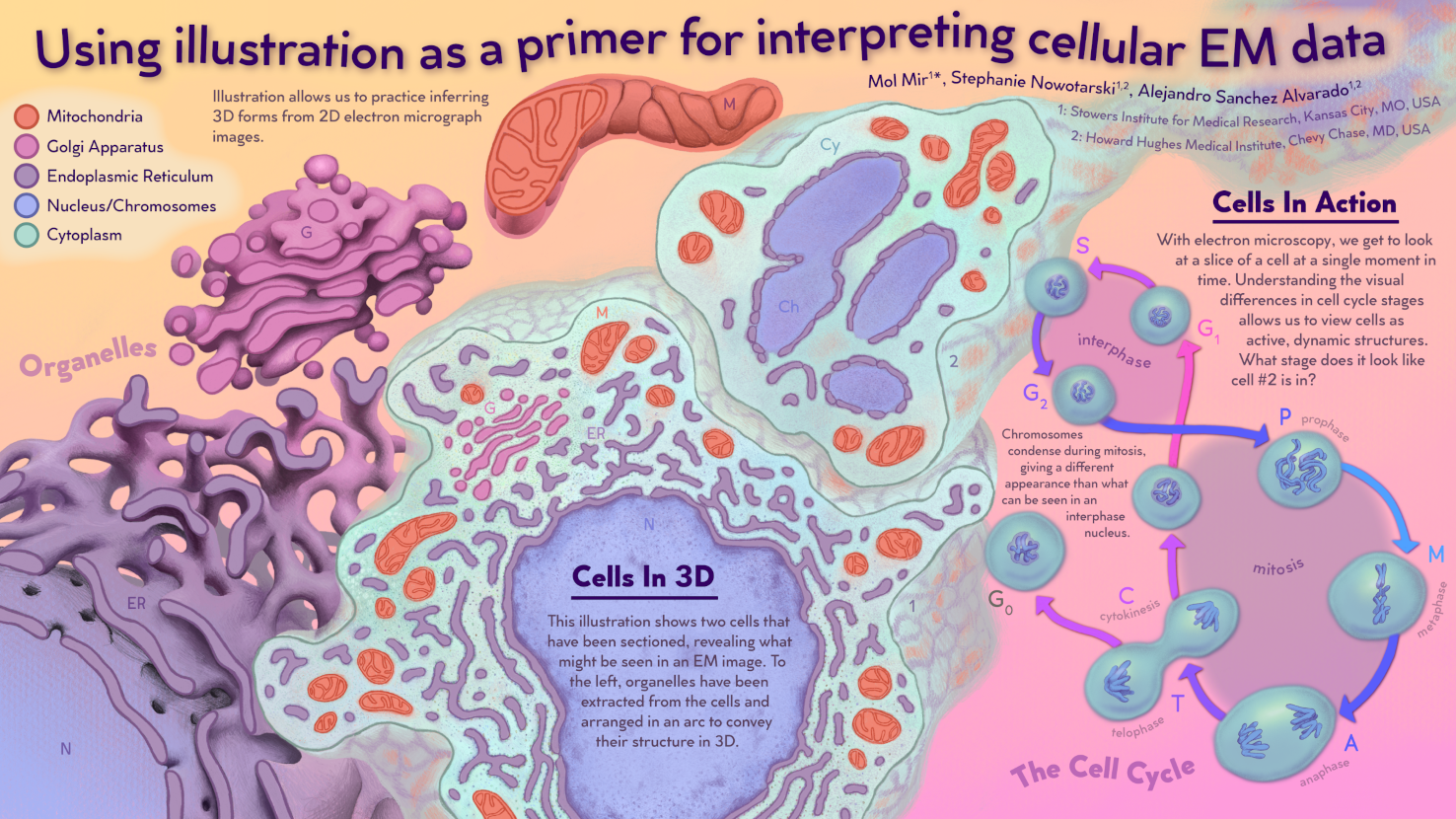
You won the Best Scientific Poster award at VIZBI 2022, can you briefly explain the poster you presented?
My VIZBI poster was an exploration of how I can prepare people – both scientists & non-scientists – to look at EM data. In my work with EM, I have consistently run into the problem that this is a form of microscopy that many people are not used to interpreting. When we look at EM images, we are seeing a single slice of a 3D sample that is also a snapshot of a point in time. Cells are dynamic, living, moving structures. They are differentiating and doing work. They are also filled with organelles that take on complex 3D forms that can be difficult to interpret from a single image without some practice. It takes some explanation for people to understand this when they are looking at in a static grayscale image.
What are your next steps?
I received some excellent feedback during my poster session at VIZBI that I’m excited to be moving forward with! My poster this year was an expansion of a spread from the book I am working on to describe planarian cells in electron microscopy. The most valuable thing I learned in my undergrad is the importance of audience testing my work. I’ll be putting together more spreads with information similar to my poster and sharing with members of my intended audience to get their feedback. I want to make learning about this subject as easy as possible. The words “planarian” and “electron microscopy” can seem pretty intimidating, but in all reality, I’m not presenting material that is beyond anyone’s understanding. It’s extremely important to me to find ways to include all people in my work, no matter their training or experience.
You can see more of Mol’s work by visiting molmir.com
Leonora Martínez-Nuñez Postdoctoral Associate, University of Massachusetts Medical School.
Can you tell us a bit about yourself and your research interests?
I obtained my Doctorate in Life Sciences in Mexico, where I studied Filamentous fungi, specifically proteins of the cell wall of Neurospora crassa. After grad school, I got a postdoc position at UMAss Medical School in the laboratory of Dr. Mary Munson, where I’ve been working with the Exocyst complex, in yeast, and other interesting organisms. My research interest has always been related to membrane trafficking, vesicle delivery, and exocytosis. At the Munson lab, we studied membrane trafficking using structural biology and biochemistry approaches to understand how exocytosis and vesicle delivery is regulated. My research is focused on the Exocyst complex, a multisubunit tethering complex with a major role in the last step of vesicle delivery during exocytosis. The main goal of my research is to understand how the complex regulates its function and its multiple interactions with a plethora of proteins, including SNAREs, at exocytic sites. Additionally, I’ve always been interested in illustration and creating scientific visuals to help disseminate scientific knowledge in creative ways.
What first interested you in this field of research?
I’m not sure when I was first interested in this field, I can only think of me being very curious about anything related to membranes and vesicle accumulation. During my Doctorate, I studied this from a cell biology approach, how fungal cells are shaped the way they are. In N. crassa, the model organism I studied, there is a massive accumulation of secretory vesicles at the tip carrying cell wall primer material. Many fungal cells present this apical accumulation of vesicles, but there are some differences across organisms; so I was curious and wanted to learn more about the molecular machines involved in vesicle delivery.
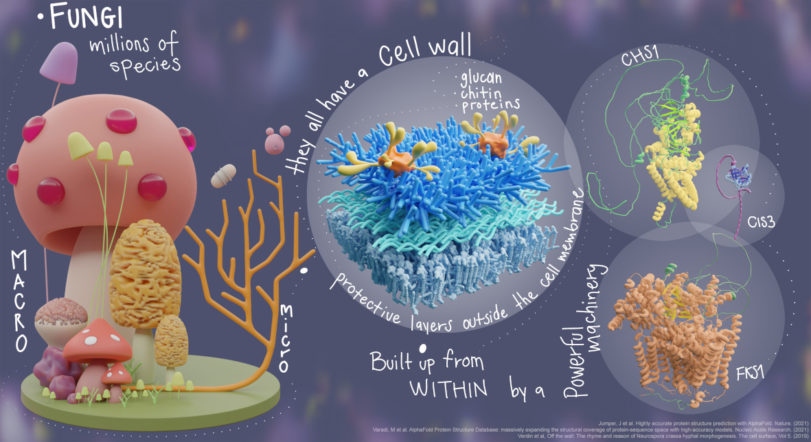
You won the Art & Biology prize at VIZBI 2022, can you briefly explain what you presented?
The poster I presented a VIZBI 2022 was actually not related to my current research, it was more of a whimsical take on what I used to work with, in combination with some protein visualization tools I’ve learned to use during my postdoc. I showed a diversity of mushrooms and microscopic fungi unified by the presence of a cell wall. Which is a protective layer that’s built from witing the fungal cells and extruded to the exterior. All fungal cell walls are mostly composed of polysaccharides and proteins embedded in a matrix-like structure on top of the cell membrane. The cell wall assembly is an orchestrated process, where multiple proteins, or powerful molecular machines, have a primary role in building. I wanted to create something that was visually appealing for a younger audience, even though VIZBI is an academic conference, and most likely no younger kids will be looking at it. My idea was to create something not too formal, with bits of information that perhaps will sparkle the audience’s interest in fungal biology.
What are your next steps?
My current research is more focused on studying Exocytosis regulation and trying to understand how all the proteins involved, actually, take part in the process. I’m currently working on building a battery of mutants that would allow me to understand how phosphorylation is part of the regulation process. However, I’m building a stronger set of skills as a scientific illustrator, and honestly, I believe that’s a better call for myself. A field where I can merge my scientific knowledge and my expertise in different scientific areas, with my interest in digital illustration and skills in 3D modelling, but of course, I feel like I’m still learning. My plan is to transition out of the wet lab in the future and find an opportunity where I can do scientific visualization/illustration for an institution perhaps. I’d say I’m open to future opportunities. Being awarded the Best poster in the Art and Biology category at VIZBI 2022, definitely helped me gain visibility, more people are aware of what I can do. This opened the possibility of potential collaborations with other scientists.
You can follow more of Leonora’s work @ leonoramartinez.com/3dscientificillustration and on Twitter (radiant_mol)/Instagram (radiant_molecules)